Ann Rehabil Med.
2012 Dec;36(6):893-896.
Extensive Intramuscular Venous Malformation in the Lower Extremity
- Affiliations
-
- 1Department of Physical Medicine & Rehabilitation, Korea University College of Medicine, Ansan 425-707, Korea. rmkdh@korea.ac.kr
Abstract
- Typical venous malformations are easily diagnosed by skin color changes, focal edema or pain. Venous malformation in the skeletal muscles, however, has the potential to be missed because their involved sites are invisible and the disease is rare. In addition, the symptoms of intramuscular venous malformation overlaps with myofascial pain syndrome or muscle strain. Most venous malformation cases have reported a focal lesion involved in one or adjacent muscles. In contrast, we have experienced a case of intramuscular venous malformation that involved a large number of muscles in a lower extremity extensively.
Keyword
MeSH Terms
Figure
-
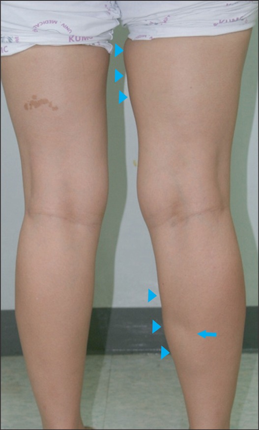
Fig. 1 Mild swelling is exhibited on the medial side of the right thigh and calf (arrow heads) and dimpling on the right medial calf area (arrow).
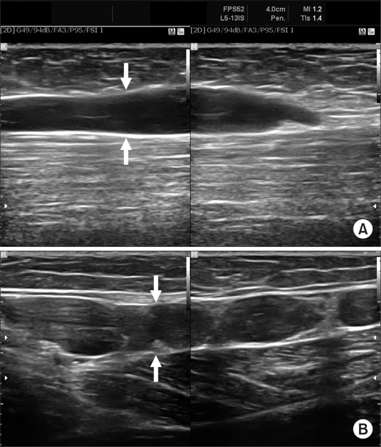
Fig. 2 Longitudinal ultrasonograms of the right medial thigh (A) and calf (B) demonstrated dilated intramuscular veins (arrows).
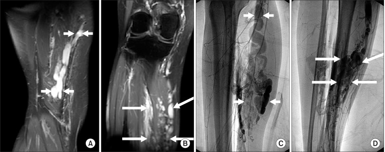
Fig. 3 Magnetic resonance images (A, B) and Venography (C, D) of the right lower extremity exhibit multiple tortous engorged venous structures mainly involving the hamstring muscles (short arrows, A, C) and soleus and gastrocnemius medial head (long arrows, B, D).
Reference
-
1. Mulliken JB, Glowacki J. Hemangiomas and vascular malformations in infants and children: a classification based on endothelial characteristics. Plast Reconstr Surg. 1982; 69:412–422. PMID: 7063565.2. Dubois J, Soulez G, Oliva VL, Berthiaume MJ, Lapierre C, Therasse E. Soft-tissue venous malformations in adult patients: imaging and therapeutic issues. Radiographics. 2001; 21:1519–1531. PMID: 11706222.
Article3. Trop I, Dubois J, Guibaud L, Grignon A, Patriquin H, McCuaig C, Garel LA. Soft-tissue venous malformations in pediatric and young adult patients: diagnosis with Doppler US. Radiology. 1999; 212:841–845. PMID: 10478255.
Article4. Hein KD, Mulliken JB, Kozakewich HP, Upton J, Burrows PE. Venous malformations of skeletal muscle. Plast Reconstr Surg. 2002; 110:1625–1635. PMID: 12447041.
Article5. Choi DJ, Alomari AI, Chaudry G, Orbach DB. Neurointerventional management of low-flow vascular malformations of the head and neck. Neuroimaging Clin N Am. 2009; 19:199–218. PMID: 19442906.
Article
- Full Text Links
-
- Actions
-
Cited
- CITED
-
- Close
- Share
-
- Similar articles
-
- Correction: Extensive Intramuscular Venous Malformation in the Lower Extremity
- Ultrasonography of the lower extremity veins: anatomy and basic approach
- Angiographic Findings of Congenital Vascular Malformation in Soft Tissue
- Popliteal Fossa Pain in 24 Year-old Female
- Chronic Venous Insufficiency of Lower Extremity
