Ann Dermatol.
2016 Oct;28(5):669-670. 10.5021/ad.2016.28.5.669.
Primary Cutaneous Apocrine Carcinoma
- Affiliations
-
- 1Department of Dermatology, Kyung Hee University College of Medicine, Seoul, Korea. bellotte@hanmail.net
- KMID: 2382904
- DOI: http://doi.org/10.5021/ad.2016.28.5.669
Abstract
- No abstract available.
Figure
-
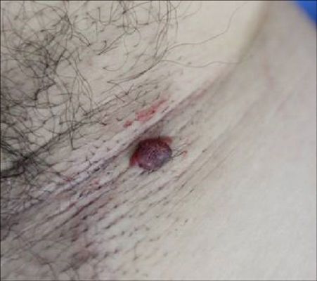
Fig. 1 The 1.2×1.0 cm sized flesh to reddish colored pedunculated nodule on the right axilla.

Fig. 2 (A) Scanning view of the specimen. (B) Specimen showed a well-to-moderately differentiated adenocarcinoma located in the dermis containing ductal and glandular structures with apocrine features (H&E, ×100). (C) Dense cellular nests were composed of cells with abundant eosinophilic cytoplasm, vesicular nuclei and central large nucleoli. Some lumen showed decapitation (H&E, ×200).
Reference
-
1. Paties C, Taccagni GL, Papotti M, Valente G, Zangrandi A, Aloi F. Apocrine carcinoma of the skin. A clinicopathologic, immunocytochemical, and ultrastructural study. Cancer. 1993; 71:375–381.
Article2. Pucevich B, Catinchi-Jaime S, Ho J, Jukic DM. Invasive primary ductal apocrine adenocarcinoma of axilla: a case report with immunohistochemical profiling and a review of literature. Dermatol Online J. 2008; 14:5.
Article3. Chamberlain RS, Huber K, White JC, Travaglino-Parda R. Apocrine gland carcinoma of the axilla: review of the literature and recommendations for treatment. Am J Clin Oncol. 1999; 22:131–135.4. Horn RC. Malignant papillary cystadenoma of sweat glands with metastases to the regional lymph nodes. Surgery. 1944; 16:348–355.5. Jun IS, Haw CR, Kim NI. Case reports: apocrine gland carcinoma. Ann Dermatol. 1996; 8:253–256.
- Full Text Links
-
- Actions
-
Cited
- CITED
-
- Close
- Share
-
- Similar articles
-
- Primary Cutaneous Mucinous Carcinoma with Extramammary Paget’s Disease: Eccrine or Apocrine?
- Primary Cutaneous Apocrine Carcinoma and Syringocystadenoma Papilliferum Arising in Nevus Sebaceus: A Case Report and Review of the Literature
- Large cutaneous apocrine carcinoma occurring on right thigh aggravated after moxa treatment
- A Case of Nevus Sebaceus associated with Tubular Apocrine Adenoma and Apocrine Poroma
- Apocrine Gland Carcinoma
