From Exoscope into the Next Generation
- Affiliations
-
- 1Department of Neurosurgery, Center for Neurological Diseases, Niigata Medical Center, Niigata, Japan. nishiken@d4.dion.ne.jp
- KMID: 2382762
- DOI: http://doi.org/10.3340/jkns.2017.0202.003
Abstract
- An exoscope, high-definition video telescope operating monitor system to perform microsurgery has recently been proposed an alternative to the operating microscope. It enables surgeons to complete the operation assistance by visualizing magnified images on a display. The strong points of exoscope are the wide field of view and deep focus. It minimized the need for repositioning and refocusing during the procedure. On the other hand, limitation of magnifying object was an emphasizing weak point. The procedures are performed under 2D motion images with a visual perception through dynamic cue and stereoscopically viewing corresponding to the motion parallax. Nevertheless, stereopsis is required to improve hand and eye coordination for high precision works. Consequently novel 3D high-definition operating scopes with various mechanical designs have been developed according to recent high-tech innovations in a digital surgical technology. It will set the stage for the next generation in digital image based neurosurgery.
MeSH Terms
Figure
-

Fig. 1 Operating theater during exoscopic surgery. Setting VITOM at interval of 25 cm from the operative field allows abundant space for manipulation. Two surgeons operate with watching two displays placed face to face. VITOM: the video telescope operating monitor.
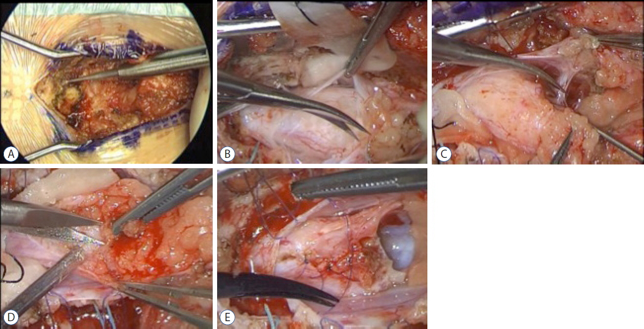
Fig. 2 An illustrative case of exoscopic surgery in pediatric spinal lipoma. A: Drilling the vertebral arches. B: Cutting dura. C: Separation of adherent arachnoid. D: Resection of lipoma along the white plane. E: Forming circular arc by sawing the pia matter.
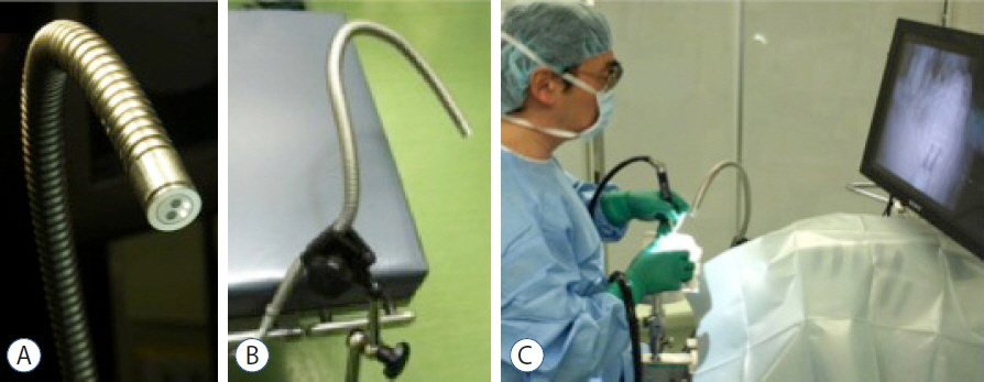
Fig. 3 3D-Eye-Flex. A: Two mini-charge coupled devices at the distal end of the scope attached to a flexible bellows. B: 3D-Eye-Flex can be fixed to an operating table. C: A circular polarizing filter system is used for the 3D display to deliver life-like images. Surgeons are able to tilt their head and examine the image with wearing special lightweight glasses.
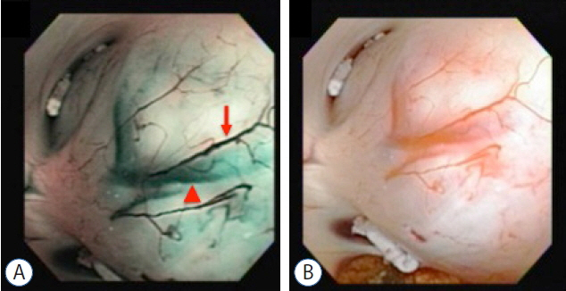
Fig. 4 A: Videoscopic findings of thalamic glioma with narrow-band imaging. An optical color separation filter narrows the bandwidth for spectral transmittance and lets only two narrow wavelengths through. These two specific wavelengths are strongly absorbed by hemoglobin. The shorter wavelengths of 415 nm penetrates only superficial layer, absorbed by capillary vessels in the surface and shows up brownish (arrow). While the longer, 540 nm light penetrates deeper, absorbed by blood vessels located subependymal layer and appears cyan on the narrow-band imaging (arrowhead). B: A standard imaging view of the same object with videoscope.
Cited by 3 articles
-
Preface : Invited Issue Editor Professor Dachling Pang and the Changing Concepts in Spinal Dysraphism during the Last Two Decades
Chae-Yong Kim, Seung-Ki Kim
J Korean Neurosurg Soc. 2020;63(3):269-271. doi: 10.3340/jkns.2020.0064.Evaluation of 3-Dimensional Exoscopes in Brain Tumor Surgery
Wan-Soo Yoon, Hyoung-Woo Lho, Dong-Sup Chung
J Korean Neurosurg Soc. 2021;64(2):289-296. doi: 10.3340/jkns.2020.0199.Assessment and Comparison of Three Dimensional Exoscopes for Near-Infrared Fluorescence-Guided Surgery Using Second-Window Indocyanine-Green
Steve S. Cho, Clare W. Teng, Emma De Ravin, Yash B. Singh, John Y.K. Lee
J Korean Neurosurg Soc. 2022;65(4):572-581. doi: 10.3340/jkns.2021.0202.
Reference
-
References
1. Hopf NJ, Perneczky A. Endoscopic neurosurgery and endoscope-assisted microneurosurgery for the treatment of intracranial cysts. Neurosurgery. 43:1330–1336. discussion 1336–1337. 1998.
Article2. Hopf NJ, Kurucz P, Reisch R. Three-dimensional HD endoscopy – first experiences with the Einstein Vision system in neurosurgery. Innovative Neurosurgery. 1:125–131. 2013.
Article3. Inoue D, Yoshimoto K, Uemura M, Yoshida M, Ohuchida K, Kenmotsu H, et al. Three-dimensional high-definition neuroendoscopic surgery: a controlled comparative laboratory study with two-dimensional endoscopy and clinical application. J Neurol Surg A Cent Eur Neurosurg. 74:357–365. 2013.
Article4. Mamelak AN, Danielpour M, Black KL, Hagike M, Berci G. A high-definition exoscope system for neurosurgery and other microsurgical disciplines: preliminary report. Surg Innov. 15:38–46. 2008.
Article5. Mamelak AN, Nobuto T, Berci G. Initial clinical experience with a high-definition exoscope system for microneurosurgery. Neurosurgery. 67:476–483. 2010.
Article6. Nishiyama K, Natori Y, Oka K. A novel three-dimensional and high-definition flexible scope. Acta Neurochir (Wien). 156:1245–1249. 2014.
Article7. Oka K. Introduction of the videoscope in neurosurgery. Neurosurgery. 62(5 Suppl 2):ONS337–ONS340. discussion ONS341. 2008.
Article8. Shirzadi A, Mukherjee D, Drazin DG, Paff M, Perri B, Mamelak AN, et al. Use of the video telescope operating monitor (VITOM) as an alternative to the operating microscope in spine surgery. Spine (Phila Pa 1976). 37:E1517–E1523. 2012.
Article9. Tabaee A, Anand VK, Fraser JF, Brown SM, Singh A, Schwartz TH. Three-dimensional endoscopic pituitary surgery. Neurosurgery. 64(5 Suppl 2):288–293. discussion 294–295. 2009.
Article10. Yoshimoto K, Mukae N, Kuga D, Inoue D, Hashizume M, Iihara K. Dual optical channel three-dimensional neuroendoscopy: Clinical application as an assistive technique in endoscopic endonasal surgery. Interdiscip Neurosurg. 6:45–50. 2016.
Article
- Full Text Links
-
- Actions
-
Cited
- CITED
-
- Close
- Share
-
- Similar articles
-
- Evaluation of 3-Dimensional Exoscopes in Brain Tumor Surgery
- The Exoscope versus operating microscope in microvascular surgery: A simulation non-inferiority trial
- Early Experience, Setup, Learning Curve, Benefits, and Complications Associated with Exoscope and Three-Dimensional 4K Hybrid Digital Visualizations in Minimally Invasive Spine Surgery
- Assessment and Comparison of Three Dimensional Exoscopes for Near-Infrared Fluorescence-Guided Surgery Using Second-Window Indocyanine-Green
- Comparison of Radiation Dose and Image Quality between the 2nd Generation and 3rd Generation Dual-Source Single-Energy and Dual-Source Dual-Energy CT of the Abdomen
