Evaluation of 3-Dimensional Exoscopes in Brain Tumor Surgery
- Affiliations
-
- 1Department of Neurosurgery, Incheon St. Mary’s Hospital, College of Medicine, The Catholic University of Korea, Incheon, Korea
- KMID: 2513860
- DOI: http://doi.org/10.3340/jkns.2020.0199
Abstract
Objective
: Though the operating microscope (OM) has been the standard optical system in neurosurgery, a new technology called three-dimensional (3D) exoscope has emerged as an alternative. Herein, two types of 3D exoscopes for brain tumor surgery are presented. In addition, the advantages and limitations compared with the OM are discussed.
Methods
: In the present study, 3D exoscope VOMS-100 or VITOM 3D was used in 11 patients with brain tumor who underwent surgical resection; the Kinevo 900 OM was used only in emergency. After completion of all surgeries, the participants were surveyed with a questionnaire regarding video image quality on the display monitor, handling of equipment, ergonomics, educational usefulness, 3D glasses, and expectation as a substitute for the OM.
Results
: Among 11 patients, nine patients underwent neurosurgical resection with only 3D exoscope; however, two patients required additional aid with the OM due to difficulty in hemostasis. Regarding video image quality, VITOM 3D was mostly equivalent to the OM, but VOMS-100 was not. However, both 3D exoscopes showed advantages in accessibility of instruments in the surgical field and occupied less space in the operating theater. Differences in ergonomics and educational usefulness between the exoscopes were not reported. Respondents did not experience discomfort in wearing 3D glasses and thought the exoscopes could be currently, and in the future, used as a substitute for the OM.
Conclusion
: Although many neurosurgeons are not familiar with 3D exoscopes, they have advantages compared with the OM and similar image quality. Exoscopes could be a substitute for OM in the future if some limitations are overcome.
Figure
-

Fig. 1. Operating room during exoscopic surgery using VITOM 3D. The 3D monitor is displayed in front of surgeons. Surgeons can see the surgical field with the naked eye due to the long working distance of the exoscope and can watch the magnified image on the monitor while wearing 3D glasses.
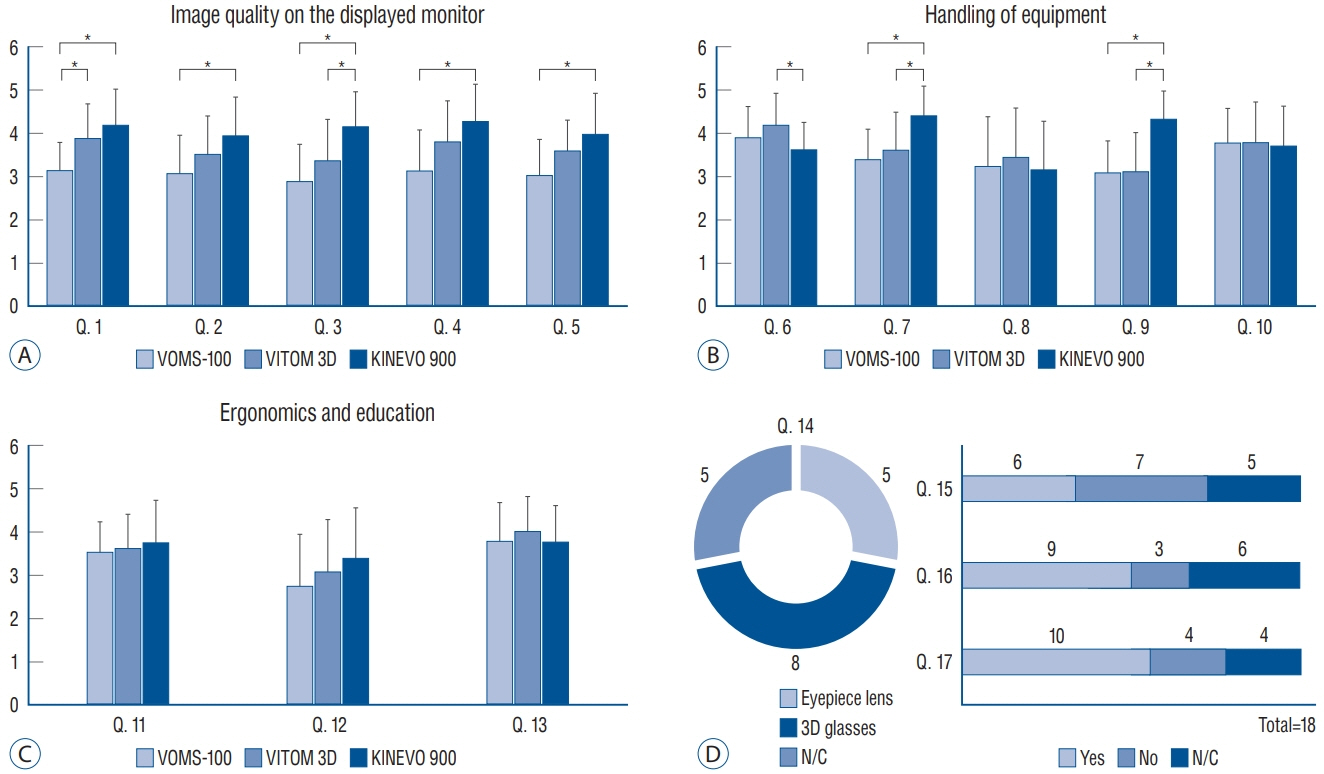
Fig. 2. Results of questionnaire regarding the experience of using exoscopes (VOMS-100 or VITOM 3D) compared with the OM (Kinevo 900). A : Questions regarding image quality on the display monitor. VITOM 3D was equivalent to Kinevo 900 except for quality of the magnified image (Q. 3). However, VOMS-100 was inferior to Kinevo 900 in all aspects (Q. 1–5). B : Questions regarding handling of equipment. Exoscopes were inferior in focusing on the surgical field (Q. 7). However, exoscopes were superior in accessibility of instruments in the surgical field (Q. 6) and amount of space occupied in the operating theater (Q. 9). C : Questions regarding ergonomics and educational usefulness. Difference was not observed between exoscope and OM. D : Questions regarding 3D glasses and expectations. Discomfort in wearing the 3D glasses was not reported, and respondents had high expectations regarding substitution of exoscopes for the OM. *p<0.05. Q : question, N/C : no choice, OM : operating microscope.
Reference
-
References
1. Beez T, Munoz-Bendix C, Beseoglu K, Steiger HJ, Ahmadi SA. First clinical applications of a high-definition three-dimensional exoscope in pediatric neurosurgery. Cureus. 10:e2108. 2018.
Article2. Gildenberg PL, Labuz J. Stereotactic craniotomy with the exoscope. Stereotact Funct Neurosurg. 68:64–71. 1997.
Article3. Gildenberg PL, Labuz J. Use of a volumetric target for image-guided surgery. Neurosurgery. 59:651–659. 2006.
Article4. Gildenberg PL, Ledoux R, Cosman E, Labuz J. The exoscope--a framebased video/graphics system for intraoperative guidance of surgical resection. Stereotact Funct Neurosurg. 63:23–25. 1994.
Article5. Jacobson JH 2nd, Wallman LJ, Schumacher GA, Flanagan M, Suarez EL, Donaghy RM. Microsurgery as an aid to middle cerebral artery endarterectomy. J Neurosurg. 19:108–115. 1962.
Article6. KARL STORZ. Highlights MICROSCOPY. Available at : https://www.karlstorz.com/cps/rde/xbcr/karlstorz_assets/ASSETS/3620832.pdf.7. Kuchta J, Simons P. Spinal neurosurgery with the head-mounted “varioscope” microscope. Cent Eur Neurosurg. 70:98–100. 2009.
Article8. Mamelak AN, Danielpour M, Black KL, Hagike M, Berci G. A high-definition exoscope system for neurosurgery and other microsurgical disciplines: preliminary report. Surg Innov. 15:38–46. 2008.
Article9. Mamelak AN, Nobuto T, Berci G. Initial clinical experience with a highdefinition exoscope system for microneurosurgery. Neurosurgery. 67:476–483. 2010.
Article10. Nishiyama K. From exoscope into the next generation. J Korean Neurosurg Soc. 60:289–293. 2017.
Article11. Oertel JM, Burkhardt BW. Vitom-3D for exoscopic neurosurgery: initial experience in cranial and spinal procedures. World Neurosurg. 105:153–162. 2017.
Article12. Ricciardi L, Chaichana KL, Cardia A, Stifano V, Rossini Z, Olivi A, et al. The exoscope in neurosurgery: an innovative “point of view”. A systematic review of the technical, surgical, and educational aspects. World Neurosurg. 124:136–144. 2019.
Article13. Rossini Z, Cardia A, Milani D, Lasio GB, Fornari M, D’Angelo V. VITOM 3D: preliminary experience in cranial surgery. World Neurosurg. 107:663–668. 2017.
Article14. SOMETECH Co., Ltd. 3D surgical video microscope (model : VOMS-100). Available at : http://www.sometech.com/bbs/product_view.php?pmode=view&idx=3&cat_no=2&offset=.15. Uluç K, Kujoth GC, Başkaya MK. Operating microscopes: past, present, and future. Neurosurg Focus. 27:E4. 2009.
Article
- Full Text Links
-
- Actions
-
Cited
- CITED
-
- Close
- Share
-
- Similar articles
-
- Assessment and Comparison of Three Dimensional Exoscopes for Near-Infrared Fluorescence-Guided Surgery Using Second-Window Indocyanine-Green
- Usefulness of Two-dimensioanl CT & Three-dimensional CT in Blow-out Fracture
- Development of Experimental 9L Gliosarcoma Rat Brain Tumor Model
- Evaluation immunocompetence in the patients with the brain tumor
- The Usefulness of Three Dimensional Reconstruction Imaging using Spiral CT: an Experience in the Bilateral Wilms` Tumor Surgery
