J Adv Prosthodont.
2017 Jun;9(3):176-181. 10.4047/jap.2017.9.3.176.
Evaluation of marginal and internal gaps of Ni-Cr and Co-Cr alloy copings manufactured by microstereolithography
- Affiliations
-
- 1Department of Dental Laboratory Science and Engineering, College of Health Science, Korea University, Seoul, Republic of Korea. kuc2842@korea.ac.kr
- 2Department of Dental Laboratory Science and Engineering, College of Health Science & Department of Public Health Sciences, Graduate School & BK21+ Program in Public Health Sciences, Korea University, Seoul, Republic of Korea.
- KMID: 2382603
- DOI: http://doi.org/10.4047/jap.2017.9.3.176
Abstract
- PURPOSE
The purpose of this study was to evaluate the marginal and internal gaps of Ni-Cr and Co-Cr copings, fabricated using the dental µ-SLA system.
MATERIALS AND METHODS
Ten study dies were made using a two-step silicone impression with a dental stone (type IV) from the master die of a tooth. Ni-Cr (NC group) and Co-Cr (CC group) alloy copings were designed using a dental scanner, CAD software, resin coping, and casting process. In addition, 10 Ni-Cr alloy copings were manufactured using the lost-wax technique (LW group). The marginal and internal gaps in the 3 groups were measured using a digital microscope (160 ×) with the silicone replica technique, and the obtained data were analyzed using the non-parametric Kruskal-Wallis H test. Post-hoc comparisons were performed using Bonferroni-corrected Mann-Whitney U tests (α=.05).
RESULTS
The mean (±standard deviation) values of the marginal, chamfer, axial wall, and occlusal gaps in the 3 groups were as follows: 81.5±73.8, 98.1±76.1, 87.1±44.8, and 146.8±78.7 µm in the LW group; 76.8±48.0, 141.7±57.1, 80.7±47.5, and 194.69±63.8 µm in the NC group; and 124.2±52.0, 199.5±71.0, 67.1±37.6, and 244.5±58.9 µm in the CC group.
CONCLUSION
The marginal gap in the LW and NC groups were clinically acceptable. Further improvement is needed for CC group to be used clinical practice.
Keyword
Figure
-

Fig. 1 Schematic diagram of the experiment.
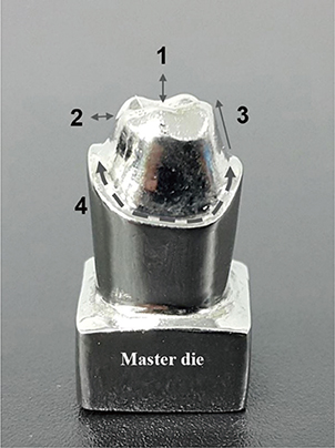
Fig. 2 Master die fabrication in casting.
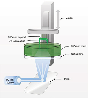
Fig. 3 Schematic diagram of the µ-SLA system principle.

Fig. 4 The silicone replica technique.

Fig. 5 Measurement point areas of marginal and internal gaps.
Reference
-
1. Konstantoulakis E, Nakajima H, Woody RD, Miller AW. Marginal fit and surface roughness of crowns made with an accelerated casting technique. J Prosthet Dent. 1998; 80:337–345.2. Tao J, Yoda M, Kimura K, Okuno O. Fit of metal ceramic crowns cast in Au-1.6 wt% Ti alloy for different abutment finish line curvature. Dent Mater. 2006; 22:397–404.3. Ucar Y, Akova T, Akyil MS, Brantley WA. Internal fit evaluation of crowns prepared using a new dental crown fabrication technique: laser-sintered Co-Cr crowns. J Prosthet Dent. 2009; 102:253–259.4. Duret F, Blouin JL, Duret B. CAD-CAM in dentistry. J Am Dent Assoc. 1988; 117:715–720.5. Miyazaki T, Hotta Y, Kunii J, Kuriyama S, Tamaki Y. A review of dental CAD/CAM: current status and future perspectives from 20 years of experience. Dent Mater J. 2009; 28:44–56.6. Davis DR. Limiting wax pattern distortion caused by setting expansion. J Prosthet Dent. 1987; 58:229–234.7. van Noort R. The future of dental devices is digital. Dent Mater. 2012; 28:3–12.8. Kim CM, Kim SR, Kim JH, Kim HY, Kim WC. Trueness of milled prostheses according to number of ball-end mill burs. J Prosthet Dent. 2016; 115:624–629.9. Örtorp A, Jönsson D, Mouhsen A, Vult von. The fit of cobalt-chromium three-unit fixed dental prostheses fabricated with four different techniques: a comparative in vitro study. Dent Mater. 2011; 27:356–363.10. Huang SH, Liu P, Mokasdar A, Hou L. Additive manufacturing and its societal impact: a literature review. Int J Adv Manuf Technol. 2013; 67:1191–1203.11. Park JY, Kim HY, Kim JH, Kim JH, Kim WC. Comparison of prosthetic models produced by traditional and additive manufacturing methods. J Adv Prosthodont. 2015; 7:294–302.12. Lee IH, Cho DW. Micro-stereolithography photopolymer solidification patterns for various laser beam exposure conditions. Int J Adv Manuf Technol. 2003; 22:410–416.13. Akova T, Ucar Y, Tukay A, Balkaya MC, Brantley WA. Comparison of the bond strength of laser-sintered and cast base metal dental alloys to porcelain. Dent Mater. 2008; 24:1400–1404.14. Kim KB, Kim WC, Kim HY, Kim JH. An evaluation of marginal fit of three-unit fixed dental prostheses fabricated by direct metal laser sintering system. Dent Mater. 2013; 29:e91–e96.15. Furst A, Radding SB. New developments in the study of metal carcinogenesis. J Environ Sci Health Part C. 1984; 2:103–133.16. Lu Y, Chen W, Ke W, Wu S. Nickel-based (Ni-Cr and Ni-Cr-Be) alloys used in dental restorations may be a potential cause for immune-mediated hypersensitivity. Med Hypotheses. 2009; 73:716–717.17. Reclaru L, Unger RE, Kirkpatrick CJ, Susz C, Eschler PY, Zuercher MH, Antoniac I, Lüthy H. Ni-Cr based dental alloys; Ni release, corrosion and biological evaluation. Mater Sci Eng C Mater Biol Appl. 2012; 32:1452–1460.18. Pradíes G, Zarauz C, Valverde A, Ferreiroa A, Martínez-Rus F. Clinical evaluation comparing the fit of all-ceramic crowns obtained from silicone and digital intraoral impressions based on wavefront sampling technology. J Dent. 2015; 43:201–208.19. Aktas G, Özcan N, Aydin DH, Şahin E, Akça K. Effect of digitizing techniques on the fit of implant-retained crowns with different antirotational abutment features. J Prosthet Dent. 2014; 111:367–372.20. Chazine M, Casucci A, Mazzoni A, Grandini S, Goracci C, Breschi L, Ferrari M. Interfacial nanoleakage and internal cement thickness of three esthetic crown systems. Dent Mater. 2012; 28:1105–1111.21. Beuer F, Aggstaller H, Edelhoff D, Gernet W, Sorensen J. Marginal and internal fits of fixed dental prostheses zirconia retainers. Dent Mater. 2009; 25:94–102.22. Karlsson S. The fit of Procera titanium crowns. An in vitro and clinical study. Acta Odontol Scand. 1993; 51:129–134.23. McLean JW, von Fraunhofer JA. The estimation of cement film thickness by an in vivo technique. Br Dent J. 1971; 131:107–111.24. Fransson B, Oilo G, Gjeitanger R. The fit of metal-ceramic crowns, a clinical study. Dent Mater. 1985; 1:197–199.25. Syrek A, Reich G, Ranftl D, Klein C, Cerny B, Brodesser J. Clinical evaluation of all-ceramic crowns fabricated from intraoral digital impressions based on the principle of active wavefront sampling. J Dent. 2010; 38:553–559.26. Boening KW, Walter MH, Reppel PD. Non-cast titanium restorations in fixed prosthodontics. J Oral Rehabil. 1992; 19:281–287.27. Jacobs MS, Windeler AS. An investigation of dental luting cement solubility as a function of the marginal gap. J Prosthet Dent. 1991; 65:436–442.28. Kokubo Y, Tsumita M, Kano T, Sakurai S, Fukushima S. Clinical marginal and internal gaps of zirconia all-ceramic crowns. J Prosthodont Res. 2011; 55:40–43.29. Elshahawy W. Marginal accuracy in casting titanium fixed partial dentures. Tanta Dent J. 2015; 12:119–123.30. Singh V, Gupta S, Bhargava A, Kaul S. Marginal accuracy of metal copings produced with different ring casting techniques: An in vitro study. Eur J Prosthodont. 2015; 3:36–41.31. Molin M, Karlsson S. The fit of gold inlays and three ceramic inlay systems. A clinical and in vitro study. Acta Odontol Scand. 1993; 51:201–206.32. Grenade C, Mainjot A, Vanheusden A. Fit of single tooth zirconia copings: comparison between various manufacturing processes. J Prosthet Dent. 2011; 105:249–255.33. de Torres EM, Rodrigues RC, de Mattos Mda G, Ribeiro RF. The effect of commercially pure titanium and alternative dental alloys on the marginal fit of one-piece cast implant frameworks. J Dent. 2007; 35:800–805.34. Hutton JE, Marshall GW. The expansion of phosphate bonded investments: Part I-Setting expansion. J Prosthet Dent. 1993; 70:121–125.
- Full Text Links
-
- Actions
-
Cited
- CITED
-
- Close
- Share
-
- Similar articles
-
- The study of tension characteristics in orthodontic wires
- Evaluation and comparison of the marginal adaptation of two different substructure materials
- Study on the effect of soldering methods on the characteristics of the Ni-Cr alloy
- Influence of the accuracy of abutment tooth preparation on the marginal adaptation of Co-Cr alloy copings fabricated with a selective laser sintering technology
- SHEAR BOND STRENGTH OF RESIN ADHESIVE CEMENT TO ENAMEL AND Ni-Cr-Be ALLOY
