Fallopian Metaplastic Papillary Tumour: An Atypical Transdifferentiation of the Tubal Epithelium?
- Affiliations
-
- 1Anatomical Pathology Division, "Dr. Manuel Gea Gonzalez" General Hospital, Mexico City, Mexico. k7nigricans@hotmail.com
- 2Pathology Unit, Mexico General Hospital, Mexico City, Mexico.
- KMID: 2381370
- DOI: http://doi.org/10.4132/jptm.2014.10.15
Abstract
- A metaplastic papillary tumor of the Fallopian tube is an extremely uncommon condition, with odd and confusing features that make it difficult to categorize as benign or borderline. Here, we summarize all the published cases to date and document the case of a 41-year-old woman diagnosed with this alteration after her last childbirth and ensuing tubal ligation. One of the tubes was bulky and filled with a caramel-like substance encircling a blurry spot. Light microscopy detailed a slender stalk covered by eosinophilic, columnar plump cells, showing atypical nuclei and focal budding. Mitotic figures were absent. The immunohistochemistry panel was positive for pan-cytokeratin, epithelial membrane antigen, cyclin D1, and hormone receptors. Additionally, a proliferation index of less than 5% was rated using Ki-67. The true nature of this tumor (reactive vs neoplastic) is uncertain. Nonetheless, its association with pregnancy suggests an adaptive change, likely similar to the atypical transdifferentiation proposed for Arias-Stella reaction.
Keyword
MeSH Terms
Figure
-

Fig. 1. Gross features/oxyphil metaplastic components. (A) Tubal ligation specimen. (B) Transverse cut surface. A crystallized syrup-like substance with a striking resemblance to a regional candy known as «acitrón» fills the lumen; there is also a left marginal blur with a snowflakelike appearance. The left column displays whole-mount sections stained with hematoxylin and eosin, periodic acid–Schiff, Alcian blue, and mucicarmine. (C) Panoramic photomicrograph showing a peninsular papillary framework in a lake of extracellular mucin. The stalk has a loose stroma and contains a small number of inflammatory cells (lymphocytes and neutrophils). (D) High magnification photomicrograph of the oncocytic epithelium. There is mild stratification and cell budding.

Fig. 2. Mucinous metaplastic components. (A) Some amphophilic mucin-filled cysts are noticeable beneath the epithelium (blue arrows). (B) Periodic acid−Schiff. (C) Alcian blue. (D) Mayer's mucicarmine.
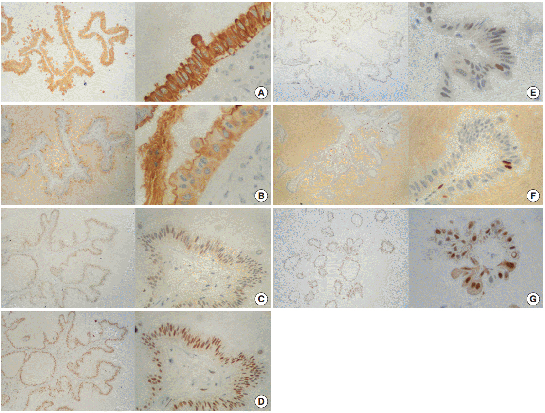
Fig. 3. Immunohistochemistry panel. (A) Cytokeratin AE1/AE3 (+++/+++) in 100% of cells. (B) Epithelial membrane antigen (+++/+++) in 100% of cells. (C) Estrogen receptor (++/+++) in ~70% of cells. (D) Progesterone receptor (+++/+++) in ~95% of cells. (E) Androgen receptor (+/+++) in ~10% of cells. (F) Ki-67 (+++/+++) in ~3% of cells. (G) Cyclin-D1 (+++/+++) in ~90% of cells.
Reference
-
1. Saffos RO, Rhatigan RM, Scully RE. Metaplastic papillary tumor of the Fallopian tube: a distinctive lesion of pregnancy. Am J Clin Pathol. 1980; 74:232–6.2. Starr AJ, Ruffolo EH, Shenoy BV, Marston BR. Primary carcinoma of the Fallopian tube: a surprise finding in a postpartum tubal ligation. Am J Obstet Gynecol. 1978; 132:344–5.
Article3. Keeney GL, Thrasher TV. Metaplastic papillary tumor of the Fallopian tube: a case report with ultrastructure. Int J Gynecol Pathol. 1988; 7:86–92.4. Bartnik J, Powell WS, Moriber-Katz S, Amenta PS. Metaplastic papillary tumor of the Fallopian tube. Case report, immunohistochemical features, and review of the literature. Arch Pathol Lab Med. 1989; 113:545–7.5. Pang LC. Hydrosalpinx due to asymptomatic bilateral tubal pregnancies associated with metaplastic papillary tumor of the Fallopian tube. South Med J. 1999; 92:725–7.
Article6. Solomon AC, Chen PJ, LiVolsi VA. Pathologic quiz case: an incidental finding in the Fallopian tube. Fallopian tube, left, tubal ligation: metaplastic papillary tumor of Fallopian tube. Arch Pathol Lab Med. 2003; 127:e363–4.7. D’Adda T, Pizzi S, Bottarelli L, Azzoni C, Manni S, Giordano G. Metaplastic papillary tumor of the salpinx: report of a case using microsatellite analysis. Int J Gynecol Pathol. 2011; 30:532–5.8. Vang R, Wheeler JE. Diseases of the Fallopian tube and paratubal region. In : Kurman RJ, Ellenson LH, Ronnett BM, editors. Blaustein´s pathology of the female genital tract. 6th ed. New York: Springer;2011. p. 529–78.9. Arias-Stella J. The Arias-Stella reaction: facts and fancies four decades after. Adv Anat Pathol. 2002; 9:12–23.
Article10. Selman K, Kafatos FC. Transdifferentiation in the labial gland of silk moths: is DNA required for cellular metamorphosis? Cell Differ. 1974; 3:81–94.
Article11. Rabban JT, Soslow RA, Zaloudek CZ. Immunohistology of the female genital tract. In : Dabbs DJ, editor. Diagnostic immunohistochemistry: theranostic and genomic applications. 3rd ed. Philadelphia: Saunders Elsevier;2010. p. 690–762.12. Kurman RJ, Vang R, Junge J, Hannibal CG, Kjaer SK, Shih IM. Papillary tubal hyperplasia: the putative precursor of ovarian atypical proliferative (borderline) serous tumors, noninvasive implants, and endosalpingiosis. Am J Surg Pathol. 2011; 35:1605–14.13. Alvarado-Cabrero I. Pathology of the Fallopian tube and broad ligament. In : Nucci MR, Oliva E, editors. Gynecologic pathology: foundations in diagnostic pathology. Philadelphia: Churchill Linvingstone;2009. p. 331–66.14. Krasevic M, Stankovic T, Petrovic O, Smiljan-Severinski N. Serous borderline tumor of the Fallopian tube presented as hematosalpinx: a case report. BMC Cancer. 2005; 5:129.
Article15. Feeley L, Quinn CM. Columnar cell lesions of the breast. Histopathology. 2008; 52:11–9.
Article16. Walker RA, Hanby A, Pinder SE, Thomas J, Ellis IO; National Coordinating Committee for Breast Pathology Research Subgroup. Current issues in diagnostic breast pathology. J Clin Pathol. 2012; 65:771–85.
Article17. DeLellis RA, Shin SJ, Treaba DO. Immunohistology of endocrine tumors. In : Dabbs DJ, editor. Diagnostic Immunohistochemistry. Theranostic and Genomic Applications. 3rd ed. Philadelphia: Saunders Elsevier;2010. p. 291–339.
- Full Text Links
-
- Actions
-
Cited
- CITED
-
- Close
- Share
-
- Similar articles
-
- Tubal pregnancy with accessory fallopian tube: A case report
- Metaplastic Variant of the Gallbladder Adenoma: A report of a case
- Isolated Torsion of Fallopian Tubes: Detection of Pedicle at Tubal End: Two Case Report
- A case of normal pregnancy after pelviscopic salpingostomy with laser for tubal pregnancy in the single fallopian tube
- Benign Teratomas of the Fallopian Tubes: A report of two cases
