Ewing's Sarcoma/Primitive Neuroectodermal Tumor of the Uterine Corpus
- Affiliations
-
- 1Department of Pathology, Korea University Ansan Hospital, Ansan, Korea. a9604@chollian.net
- 2Department of Pathology, Korea University Anam Hospital, Seoul, Korea.
- KMID: 2381355
- DOI: http://doi.org/10.4132/jptm.2014.10.14
Abstract
- No abstract available.
MeSH Terms
Figure
-
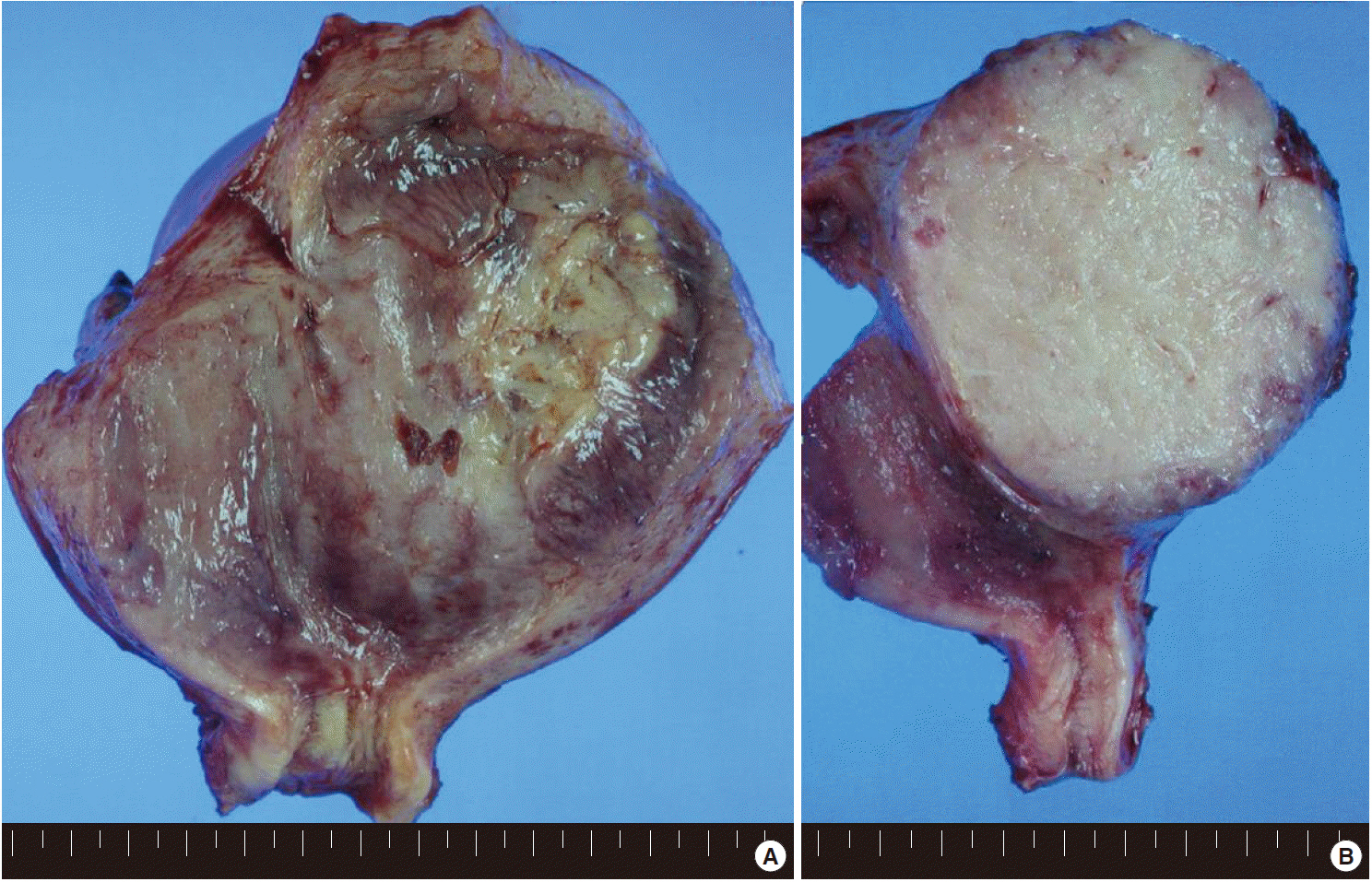
Fig. 1. (A) The uterus is slightly enlarged, measuring 6×13×8 cm. On opening the uterus, there is a mass arising from the anterior wall of the uterus, bulging out into the endometrial cavity with an area of ulceration on the endometrium. (B) The cut surface of the uterus shows an unencapsulated but relatively well-circumscribed intramural tumor, measuring 9×7.5 cm. The tumor shows a homogeneous gray-tan, solid, and fish-fleshy appearing cut surface with no conspicuous necrosis or hemorrhage. The tumor abuts the endometrium and serosal surface of the uterus.
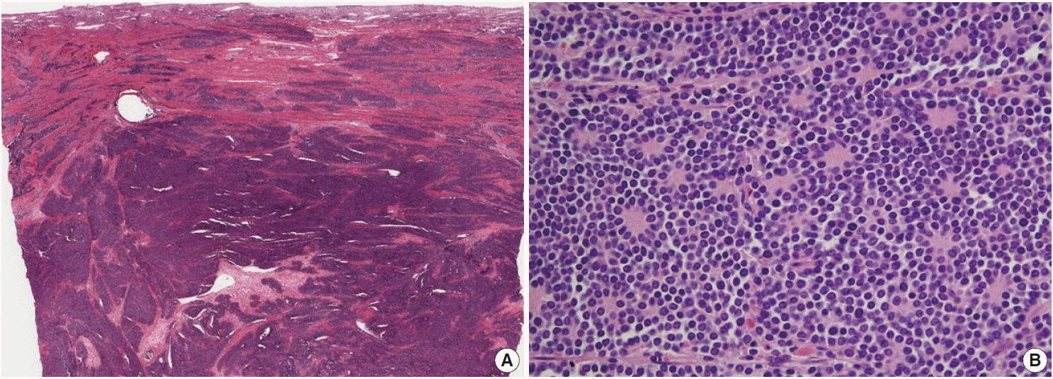
Fig. 2. (A) The tumor is composed of relatively uniform small round-to-oval neoplastic cells and arranged in a diffuse sheet or solid nesting pattern of growth with intervening fibrous septa throughout the myometrium. The tumor invades the endometrium focally but does not involve the serosal surface of the uterus. (B) The tumor cells have scant cytoplasm with an indistinct cytoplasmic border, round-to-oval nuclei of stippled chromatin pattern, and inconspicuous nucleoli. Pseudorosettes are also frequently present but no malignant glandular areas are identified within or adjacent to the tumor.
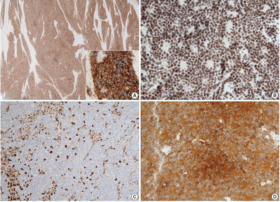
Fig. 3. The tumor cells show diffuse strong positivity for CD99 (A) and neuron-specific enolase (D) in a membrane pattern and FLI-1 (B) in a nuclear pattern. The tumor cells are focally positive for vimentin (C).
Reference
-
1. Hart MN, Earle KM. Primitive neuroectodermal tumors of the brain in children. Cancer. 1973; 32:890–7.
Article2. Masoura S, Kourtis A, Kalogiannidis I, et al. Primary primitive neuroectodermal tumor of the cervix confirmed with molecular analysis in a 23-year-old woman: a case report. Pathol Res Pract. 2012; 208:245–9.
Article3. Truong AH, Ben-David Y. The role of Fli-1 in normal cell function and malignant transformation. Oncogene. 2000; 19:6482–9.
Article4. Kleinman GM, Young RH, Scully RE. Primary neuroectodermal tumors of the ovary: a report of 25 cases. Am J Surg Pathol. 1993; 17:764–78.5. Vang R, Taubenberger JK, Mannion CM, et al. Primary vulvar and vaginal extraosseous Ewing’s sarcoma/peripheral neuroectodermal tumor: diagnostic confirmation with CD99 immunostaining and reverse transcriptase-polymerase chain reaction. Int J Gynecol Pathol. 2000; 19:103–9.
Article6. Liao X, Xin X, Lü X. Primary Ewing’s sarcoma-primitive neuroectodermal tumor of the vagina. Gynecol Oncol. 2004; 92:684–8.
Article7. Tsao AS, Roth LM, Sandler A, Hurteau JA. Cervical primitive neuroectodermal tumor. Gynecol Oncol. 2001; 83:138–42.
Article8. Ren YL, Tang XY, Li T. Ewing sarcoma-primitive neuroectodermal tumor of the uterus: a clinicopathologic, immunohistochemical and ultrastructural study of one case. Arch Gynecol Obstet. 2011; 283:1139–43.
Article9. Mhawech-Fauceglia P, Herrmann F, Penetrante R, et al. Diagnostic utility of FLI-1 monoclonal antibody and dual-colour, break-apart probe fluorescence in situ (FISH) analysis in Ewing’s sarcoma/primitive neuroectodermal tumour (EWS/PNET): a comparative study with CD99 and FLI-1 polyclonal antibodies. Histopathology. 2006; 49:569–75.10. Sandberg AA, Bridge JA. Updates on the cytogenetics and molecular genetics of bone and soft tissue tumors: desmoplastic small roundcell tumors. Cancer Genet Cytogenet. 2002; 138:1–10.
- Full Text Links
-
- Actions
-
Cited
- CITED
-
- Close
- Share
-
- Similar articles
-
- Ewing Sarcoma/Peripheral Primitive Neuroectodermal Tumor in an Adolscence, Manifested as Isolated Cervical Mass
- Double Primary Presentation of Liposarcoma and Ewing's Sarcoma: A Case Report
- A Case of a Primitive Neuroectodermal Tumor Detected from a Duodenal Metastasis
- Ewing's Sarcoma of the Lesser Sac Masquerading as a Pancreatic Tumor
- A case of extraosseous ewing's sarcoma/primitive neuroectodermal tumor of the ovary
