PHH3 as an Ancillary Mitotic Marker in Gastrointestinal Stromal Tumors
- Affiliations
-
- 1Department of Pathology and Translational Genomics, Samsung Medical Center, Sungkyunkwan University College of Medicine, Seoul, Korea. kkmkys@skku.edu
- KMID: 2381349
- DOI: http://doi.org/10.4132/jptm.2014.10.08
Abstract
- BACKGROUND
Counting mitoses is subjective and time-consuming. The adjunctive diagnostic utility of a recently reported mitotic marker, phosphohistone H3 (PHH3), was investigated in gastrointestinal stromal tumors (GISTs).
METHODS
We reviewed 77 GISTs for several proliferative indices. These included the mitotic count per 50 high power fields (HPFs), the immunohistochemical Ki-67 labeling index and the immunohistochemical PHH3 mitotic index (MI). For comparison, Spearman's rank correlation and interclass correlation coefficient were used.
RESULTS
Mitotic counts ranged from 0-138 (mean, 7.57+/-2.34) and the PHH3 MI ranged from 0-126 per 50 HPFs (mean, 9.61+/-2.27). We found a positive correlation between mitotic counts and PHH3 MI (r=0.810, p<.001). The inter-observer correlation coefficient for three participants was 0.975 for mitotic counts and 0.940 for the PHH3 MI. When using the PHH3 MI instead of mitotic counts in the Armed Forces Institute of Pathology (AFIP) stratification criteria, 10 cases were reclassified. In one patient with a mitotic count of 2 and a PHH3 MI of 6 per 50 HPFs, distant metastasis occurred.
CONCLUSIONS
In GISTs, the PHH3 MI correlated adequately with mitotic counts and can be used as a useful adjunctive to count mitotic figures efficiently.
MeSH Terms
Figure
-

Fig. 1. A comparison of hematoxylin and eosin (A, C) and phophohistone H3 (PHH3) immunohistochemistry (B, D) in mitotic detection (arrows). Mitotic figures (arrows) are easily and quickly recognized with PHH3 immunohistochemistry.

Fig. 2. Hematoxylin and eosin (H&E) (A) and phophohistone H (PHH3) immunohistochemistry (B) in a case with delayed fixation. H&E shows no mitotic figures whereas PHH3 shows three mitotic figures in the same field (arrows).
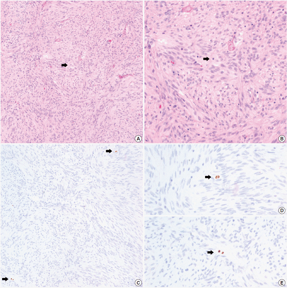
Fig. 3. Mitosis and PHH3 positive cells in a patient with distant metastasis. Hematoxylin and eosin (H&E) staining shows a single mitotic figure. (B) H&E at higher magnification. (C) Phophohistone H3 (PHH3) immunohistochemistry highlights two mitotic figures in the same field (arrows). (D, E) PHH3 at higher magnification.
Reference
-
1. Miettinen M, Lasota J. Gastrointestinal stromal tumors: review on morphology, molecular pathology, prognosis, and differential diagnosis. Arch Pathol Lab Med. 2006; 130:1466–78.
Article2. Corless CL, Fletcher JA, Heinrich MC. Biology of gastrointestinal stromal tumors. J Clin Oncol. 2004; 22:3813–25.
Article3. Franquemont DW. Differentiation and risk assessment of gastrointestinal stromal tumors. Am J Clin Pathol. 1995; 103:41–7.
Article4. Fletcher CD, Berman JJ, Corless C, et al. Diagnosis of gastrointestinal stromal tumors: a consensus approach. Hum Pathol. 2002; 33:459–65.
Article5. Miettinen M, Sobin LH, Lasota J. Gastrointestinal stromal tumors of the stomach: a clinicopathologic, immunohistochemical, and molecular genetic study of 1765 cases with long-term follow-up. Am J Surg Pathol. 2005; 29:52–68.6. Miettinen M, Makhlouf H, Sobin LH, Lasota J. Gastrointestinal stromal tumors of the jejunum and ileum: a clinicopathologic, immunohistochemical, and molecular genetic study of 906 cases before imatinib with long-term follow-up. Am J Surg Pathol. 2006; 30:477–89.7. Tapia C, Kutzner H, Mentzel T, Savic S, Baumhoer D, Glatz K. Two mitosis-specific antibodies, MPM-2 and phospho-histone H3(Ser28), allow rapid and precise determination of mitotic activity. Am J Surg Pathol. 2006; 30:83–9.8. Agaimy A. Gastrointestinal stromal tumors (GIST) from risk stratification systems to the new TNM proposal: more questions than answers? A review emphasizing the need for a standardized GIST reporting. Int J Clin Exp Pathol. 2010; 3:461–71.9. Hendzel MJ, Wei Y, Mancini MA, et al. Mitosis-specific phosphorylation of histone H3 initiates primarily within pericentromeric heterochromatin during G2 and spreads in an ordered fashion coincident with mitotic chromosome condensation. Chromosoma. 1997; 106:348–60.10. Ribalta T, McCutcheon IE, Aldape KD, Bruner JM, Fuller GN, et al. The mitosis-specific antibody anti-phosphohistone-H3 (PHH3) facilitates rapid reliable grading of meningiomas according to WHO 2000 criteria. Am J Surg Pathol. 2004; 28:1532–6.
Article11. Kim YJ, Ketter R, Steudel WI, Feiden W. Prognostic significance of the mitotic index using the mitosis marker anti-phosphohistone H3 in meningiomas. Am J Clin Pathol. 2007; 128:118–25.12. Fukushima S, Terasaki M, Sakata K, et al. Sensitivity and usefulness of anti-phosphohistone-H3 antibody immunostaining for counting mitotic figures in meningioma cases. Brain Tumor Pathol. 2009; 26:51–7.13. Bossard C, Jarry A, Colombeix C, et al. Phosphohistone H3 labelling for histoprognostic grading of breast adenocarcinomas and computer-assisted determination of mitotic index. J Clin Pathol. 2006; 59:706–10.
Article14. Nasr MR, El-Zammar O. Comparison of pHH3, Ki-67, and survivin immunoreactivity in benign and malignant melanocytic lesions. Am J Dermatopathol. 2008; 30:117–22.
Article15. Veras E, Malpica A, Deavers MT, Silva EG. Mitosis-specific marker phospho-histone H3 in the assessment of mitotic index in uterine smooth muscle tumors: a pilot study. Int J Gynecol Pathol. 2009; 28:316–21.
Article16. Tsuta K, Liu DC, Kalhor N, Wistuba II, Moran CA. Using the mitosis-specific marker anti-phosphohistone H3 to assess mitosis in pulmonary neuroendocrine carcinomas. Am J Clin Pathol. 2011; 136:252–9.
Article17. Takahashi H, Murai Y, Tsuneyama K, et al. Overexpression of phosphorylated histone H3 is an indicator of poor prognosis in gastric adenocarcinoma patients. Appl Immunohistochem Mol Morphol. 2006; 14:296–302.
Article18. Nakashima S, Shiozaki A, Ichikawa D, et al. Anti-phosphohistone H3 as an independent prognostic factor in human esophageal squamous cell carcinoma. Anticancer Res. 2013; 33:461–7.19. Kim A, Im DH, Kim K, et al. Usefulness of anti-phosphohistone H3 immunoreactivity to determine mitotic rate in gastrointestinal stromal tumors. Basic Appl Pathol. 2012; 5:91–7.
Article20. Carrillo R, Candia A, Rodriguez-Peralto JL, Caz V, et al. Prognostic significance of DNA ploidy and proliferative index (MIB-1 index) in gastrointestinal stromal tumors. Hum Pathol. 1997; 28:160–5.
Article21. Toquet C, Le Neel JC, Guillou L, et al. Elevated (> or = 10%) MIB-1 proliferative index correlates with poor outcome in gastric stromal tumor patients: a study of 35 cases. Dig Dis Sci. 2002; 47:2247–53.22. Filiz G, Yalçinkaya O, Gürel S, Yerci O, Memik F. The relationship between MIB-1 proliferative activity and mitotic index in gastrointestinal stromal tumors. Hepatogastroenterology. 2007; 54:438–41.23. Gerdes J, Lemke H, Baisch H, Wacker HH, Schwab U, Stein H. Cell cycle analysis of a cell proliferation-associated human nuclear antigen defined by the monoclonal antibody Ki-67. J Immunol. 1984; 133:1710–5.24. Juan G, Traganos F, James WM, et al. Histone H3 phosphorylation and expression of cyclins A and B1 measured in individual cells during their progression through G2 and mitosis. Cytometry. 1998; 32:71–7.
Article25. Brenner RM, Slayden OD, Rodgers WH, et al. Immunocytochemical assessment of mitotic activity with an antibody to phosphorylated histone H3 in the macaque and human endometrium. Hum Reprod. 2003; 18:1185–93.
Article26. Schimming TT, Grabellus F, Roner M, et al. pHH3 immunostaining improves interobserver agreement of mitotic index in thin melanomas. Am J Dermatopathol. 2012; 34:266–9.
Article
- Full Text Links
-
- Actions
-
Cited
- CITED
-
- Close
- Share
-
- Similar articles
-
- A Case Report of a Bleeding Duodenal Gastro-Intestinal Stromal Tumor and its Emergent Management
- Gastrointestinal Stromal Tumor with Accelerated Growth Pattern in the Stomach
- A Case of Malignant Duodenal Stromal Tumor
- A Case of Hepatic Recurrence of Low Risk Duodenal Gastrointestinal Stromal Tumor in 11 Years after Curative Resection
- A Case of Incidentally Found Esophageal Gastrointestinal Stromal Tumor
