Cancer Res Treat.
2014 Oct;46(4):415-418. 10.4143/crt.2013.068.
Pleural Metastasis as Initial Presentation of Occult Gastric Cardia Cancer: A Possible Role of PET-CT in Diagnosis
- Affiliations
-
- 1Department of Internal Medicine, Konkuk University Medical Center, Konkuk University School of Medicine, Seoul, Korea. yhcho@kuh.ac.kr
- 2Department of Pathology, Konkuk University Medical Center, Konkuk University School of Medicine, Seoul, Korea.
- 3Department of Nuclear Medicine, Konkuk University Medical Center, Konkuk University School of Medicine, Seoul, Korea.
- KMID: 2380380
- DOI: http://doi.org/10.4143/crt.2013.068
Abstract
- We report on a case of malignant pleural effusion as initial metastatic presentation of occult gastric cardia cancer in a young woman. To the best of our knowledge, this is the first report of gastric adenocarcinoma metastasized to pleura as an initial presentation. Location of cardia and signet ring cell histology may contribute to the manifestation. Utilization of positron emission tomography-computed tomography was helpful for proper diagnosis. For patients with such distinct clinical presentations, it would be appropriate to consider gastric cancer as one of the possible primary sites.
Keyword
MeSH Terms
Figure
-
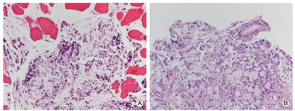
Fig. 1. (A) Pleural biopsy shows clusters of poorly differentiated carcinoma with signet ring cell features, infiltrating into the skeletal muscle fibers (H&E staining, ×200). (B) The biopsy shows clusters of poorly differentiated carcinoma with signet ring cell features and remaining gastric foveolar epithelium (upper part) (H&E staining, ×200).

Fig. 2. (A) Mild fludeoxyglucose uptake (maximum standardized uptake value [SUVmax], 4.3) seen at gastric cardia. (B) Larger and more prominent uptake (SUVmax, 8.0) seen four months later.
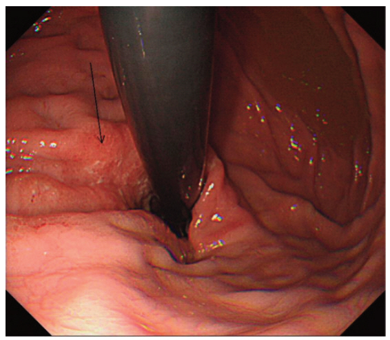
Fig. 3. Endoscopic finding of a patient with ill-defined hyperemic erosions in the cardia at retroflexion view.
Reference
-
References
1. Yoshida A, Tsuta K, Watanabe S, Sekine I, Fukayama M, Tsuda H, et al. Frequent ALK rearrangement and TTF-1/p63 co-expression in lung adenocarcinoma with signet-ring cell component. Lung Cancer. 2011; 72:309–15.
Article2. Kastelik JA. Management of malignant pleural effusion. Lung. 2013; 191:165–75.
Article3. Maeda H, Okabayashi T, Nishimori I, Sugimoto T, Namikawa T, Dabanaka K, et al. Clinicopathologic features of adenocarcinoma at the gastric cardia: is it different from distal cancer of the stomach? J Am Coll Surg. 2008; 206:306–10.
Article4. Kim JY, Lee HS, Kim N, Shin CM, Lee SH, Park YS, et al. Prevalence and clinicopathologic characteristics of gastric cardia cancer in South Korea. Helicobacter. 2012; 17:358–68.
Article5. Taghavi S, Jayarajan SN, Davey A, Willis AI. Prognostic significance of signet ring gastric cancer. J Clin Oncol. 2012; 30:3493–8.
Article6. Cheung TC, Ng FH, Chow KC, Maw CK, Ng WF. Occult gastric cancer presenting as cor pulmonale resulting from tumor cell microembolism. Am J Gastroenterol. 1997; 92:1057–9.7. Desigan G, Wang M, Wofford B, Dunn GD, Vaughan S. Occult gastric cancer manifested by progressive shortness of breath in a young adult. South Med J. 1986; 79:1173–6.
Article8. Nguyen VX, Nguyen BD, Ram PC. Occult colon cancer with initial cutaneous metastatic manifestation: PET/CT detection. Clin Nucl Med. 2012; 37:506–8.9. Patel P, Finger PT. Whole-body 18F FDG positron emission tomography/computed tomography evaluation of patients with uveal metastasis. Am J Ophthalmol. 2012; 153:661–8.
Article10. Han A, Xue J, Hu M, Zheng J, Wang X. Clinical value of 18F-FDG PET-CT in detecting primary tumor for patients with carcinoma of unknown primary. Cancer Epidemiol. 2012; 36:470–5.
Article
- Full Text Links
-
- Actions
-
Cited
- CITED
-
- Close
- Share
-
- Similar articles
-
- Late Port Site Metastasis from Occult Gall Bladder Carcinoma After Laparoscopic Cholecystectomy for Cholelithiasis: The Role of 18F-FDG PET/CT
- Occult Invasive Lobular Carcinoma of Breast Detected by Stomach Metastasis: A Case Report
- Role of F-18 FDG PET or PET/CT in the Evaluation of Gastric Cancer
- The Diagnostic Utility of PET-CT for the Preoperative Evaluation of Lymph Node Metastasis in Gastric Cancer Patients
- Effectiveness of Positron Emission Tomography in the Pre-operative Staging of Gastric Cancer
