Intense Pulsed Light Increases Hyaluronan and CD44 in Epidermal Keratinocytes and Improves Age-Related Epidermal Structure Defects in Mice
- Affiliations
-
- 1Department of Dermatology, Yonsei University College of Medicine, Seoul, Korea.
- 2Department of Applied Bioscience, CHA University, Seongnam, Korea.
- 3Institute for Clinical Research, CHA University, Seongnam, Korea.
- 4Department of Dermatology, CHA Bundang Medical Center, CHA University, Seongnam, Korea. derma97@gmail.com
- KMID: 2378549
- DOI: http://doi.org/10.5021/ad.2017.29.3.377
Abstract
- No abstract available.
Figure
-
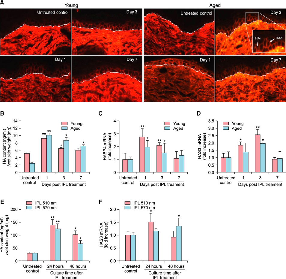
Fig. 1 Intense pulsed light (IPL) treatment increases hyaluronan (HA) production and hyaluronan synthases (HAS) 3 expression in young and old aged mouse epidermis and cultured human keratinocytes. (A) Sections of young and aged mouse skin treated with IPL were stained for HA binding protein (HABP) 4 and examined by confocal microscope. Dashed line is the dermal-epidermal junction. Magnified image shows example of accumulated HA in the intercellular (HAo) and intracellular space (HAi) (×400). After epidermal separation, HA content was measured by enzyme-linked immunosorbent assay (ELISA) (B) and the mRNA level of HABP4 (C) and HAS3 (D) was analyzed by quantitative real-time polymerase chain reaction (RT-PCR). Normal human epidermal keratinocytes (NHEK) were irradiated with IPL using either 510 nm or 570 nm cut-off filter. (E) ELISA for HA content and (F) quantitative RT-PCR analysis of HAS3 mRNA in conditioned medium from NHEK irradiated with IPL. Data represent the means±standard error. Data were analyzed by Student's t-test (*p<0.05, **p<0.01).

Fig. 2 Intense pulsed light (IPL) treatment increases CD44 expression in young and old aged mouse epidermis and cultured human keratinocytes and improves epidermal structure in aged mouse skin. (A) Immunohistochemical staining of CD44 in young and old aged mouse skin treated with IPL (×200). (B) After epidermal separation, CD44 mRNA level was analyzed by quantitative real-time polymerase chain reaction (RT-PCR). (C) CD44 mRNA level of conditioned medium from normal human epidermal keratinocytes (NHEK) irradiated with IPL using either 510 nm or 570 nm cut-off filter was analyzed by quantitative RT-PCR. Expression of differentiation marker, filaggrin (D) and proliferation marker, proliferating cell nuclear antigen (PCNA) (E) in aged mice skin treated with IPL was examined by immunohistochemical staining (×200). The stained area per epidermal area for filaggrin was measured (D) and cells with positive staining for PCNA per mm epidermal length were counted (E) by digital image analysis. Data represent the means±standard error (*p<0.05, **p<0.01).
Reference
-
1. Ghadially R, Brown BE, Sequeira-Martin SM, Feingold KR, Elias PM. The aged epidermal permeability barrier. Structural, functional, and lipid biochemical abnormalities in humans and a senescent murine model. J Clin Invest. 1995; 95:2281–2290.
Article2. Bourguignon LY, Wong G, Xia W, Man MQ, Holleran WM, Elias PM. Selective matrix (hyaluronan) interaction with CD44 and RhoGTPase signaling promotes keratinocyte functions and overcomes age-related epidermal dysfunction. J Dermatol Sci. 2013; 72:32–44.
Article3. Tzellos TG, Klagas I, Vahtsevanos K, Triaridis S, Printza A, Kyrgidis A, et al. Extrinsic ageing in the human skin is associated with alterations in the expression of hyaluronic acid and its metabolizing enzymes. Exp Dermatol. 2009; 18:1028–1035.
Article4. Oh JH, Kim YK, Jung JY, Shin JE, Chung JH. Changes in glycosaminoglycans and related proteoglycans in intrinsically aged human skin in vivo. Exp Dermatol. 2011; 20:454–456.
Article5. Goldman MP, Weiss RA, Weiss MA. Intense pulsed light as a nonablative approach to photoaging. Dermatol Surg. 2005; 31:1179–1187. discussion 1187.
Article6. Malaisse J, Bourguignon V, De Vuyst E, Lambert de Rouvroit C, Nikkels AF, Flamion B, et al. Hyaluronan metabolism in human keratinocytes and atopic dermatitis skin is driven by a balance of hyaluronan synthases 1 and 3. J Invest Dermatol. 2014; 134:2174–2182.
Article7. Sayo T, Sugiyama Y, Takahashi Y, Ozawa N, Sakai S, Ishikawa O, et al. Hyaluronan synthase 3 regulates hyaluronan synthesis in cultured human keratinocytes. J Invest Dermatol. 2002; 118:43–48.
Article8. Karvinen S, Pasonen-Seppänen S, Hyttinen JM, Pienimäki JP, Törrönen K, Jokela TA, et al. Keratinocyte growth factor stimulates migration and hyaluronan synthesis in the epidermis by activation of keratinocyte hyaluronan synthases 2 and 3. J Biol Chem. 2003; 278:49495–49504.
Article9. Pasonen-Seppänen SM, Maytin EV, Törrönen KJ, Hyttinen JM, Hascall VC, MacCallum DK, et al. All-trans retinoic acid-induced hyaluronan production and hyperplasia are partly mediated by EGFR signaling in epidermal keratinocytes. J Invest Dermatol. 2008; 128:797–807.
Article
- Full Text Links
-
- Actions
-
Cited
- CITED
-
- Close
- Share
-
- Similar articles
-
- The Effect of Intense Pulsed Light Treatment on the Epidermal Melanocytic Lesions
- Hyaluronan Oligosaccharides Improve Rosacea-Like Phenotype through Anti-Inflammatory and Epidermal Barrier-Improving Effects
- Treatment of severe erythematotelangiectatic rosacea with intense pulsed light: a case report
- Treatment of acne fulminans with intense pulsed light: a case report
- AllogeneicLymphocyte Stimulating Capacity of Contact Sensitized Epidermal Cells in Mouse
