Rehabilitation of a patient with non-syndromic partial oligodontia
- Affiliations
-
- 1Department of Prosthodontics and Research Institute of Oral Science, College of Dentistry, Gangneung-Wonju National University, Gangneung, Republic of Korea. lila@gwnu.ac.kr
- KMID: 2376851
- DOI: http://doi.org/10.4047/jap.2016.8.3.241
Abstract
- Oligodontia is defined as a congenital tooth agenesis with the absence of six or more permanent teeth. This clinical report describes a patient with non-syndromic partial oligodontia, with retained deciduous teeth and the absence of 16 permanent teeth. Anterior esthetic problems were caused by interarch tooth size discrepancy, interdental space, aberrant tooth dimensions, and the absence of centric contacts of the anterior teeth. Prosthetic restoration after orthodontic and implant treatment was performed with a multi-disciplinary team approach. Favorable functional and esthetic results were obtained using a definitive prosthesis.
Figure
-
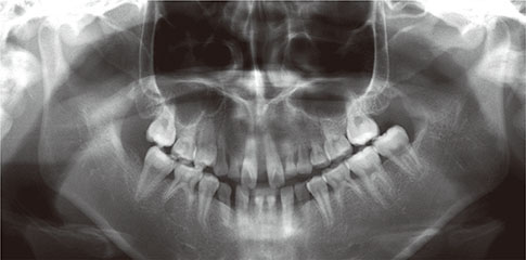
Fig. 1 Panoramic radiograph.
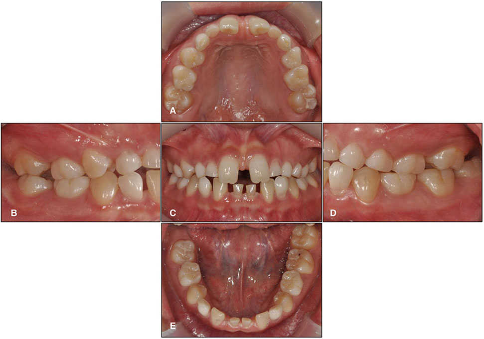
Fig. 2 Pre-operative intraoral photographs. (A) Maxillary occlusal view, (B) Right lateral view, (C) Frontal view, (D) Left lateral view, (E) Mandibular occlusal view.
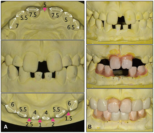
Fig. 3 (A) Anterior tooth size discrepancy. Bolton anterior ratio = 85.3%, (B) Kesling setup.
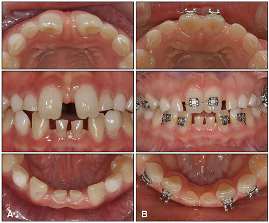
Fig. 4 Orthodontic treatment. (A) Pre-orthodontic treatment, (B) Post-orthodontic treatment.
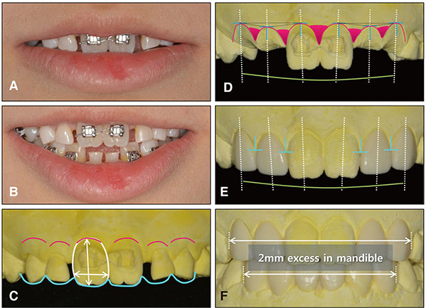
Fig. 5 Diagnostic wax-up. (A) Resting, (B) Smile, (C) Esthetic design of the planned crown. Red lines represent the ideal cervical margin. Blue lines represent the incisal edge of maxillary incisors, (D) Soft tissue contour design. Red lines represent the ideal cervical margin. Blue "T" represents the zenith of the planned anterior crowns. White lines represent the natural balance of gingival levels, (E) Diagnostic waxing after soft tissue contour design, (F) Interarch tooth size discrepany (2 mm) indicated by the Bolton anterior ratio.
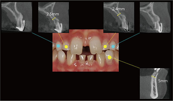
Fig. 6 CBCT images. Narrow ridge and inadequate bone width of both maxillary deciduous lateral incisor and mandibular left deciduous canine areas.

Fig. 7 Distance between the cervical margin of the crown and the planned implant position.

Fig. 8 Implant surgery procedures. (A) Bucco-lingual bone thickness of mandibular left canine region, (B) Implant placement of mandibular left canine, (C) Extraction of both maxillary deciduous canines, (D) Implant placement of both maxillary canines, (E) Provisional restorations. Note the concave emergence profile.

Fig. 9 Provisional restorations. (A) Immediately after implant placement in canine areas, (B) First provisional restoration, (C) Second provisional restoration.
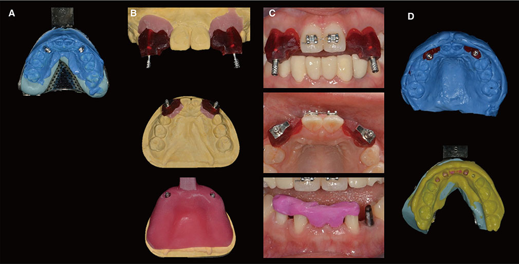
Fig. 10 Impression procedures. (A) Preliminary impression, (B) Customized pick-up impression coping and customized tray, (C) Capturing both peri-implant and pontic gingival contours, (D) Definitive impression.

Fig. 11 (A) Fabrication of zirconia coping, (B) Trial placement.
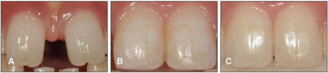
Fig. 12 Resin infiltration. (A) Pre-orthodontic treatment, (B) Post-orthodontic treatment, (C) After resin infiltration.
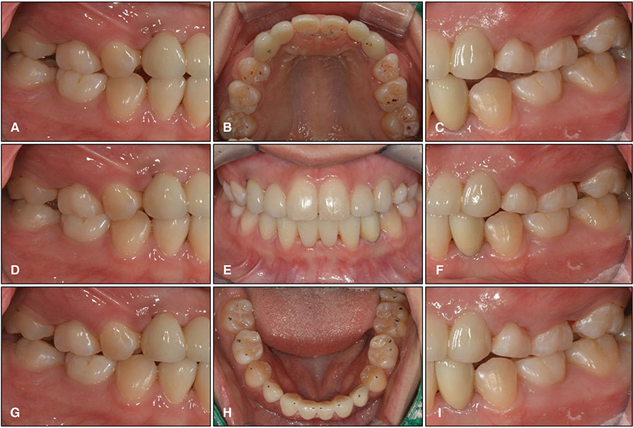
Fig. 13 Definitive prosthesis. (A) Working side during right lateral excursion, (B) Maxillary occlusal view, (C) Non-working side during right lateral excursion, (D) Left lateral view at centric occlusion, (E) Frontal view at centric occlusion, (F) Right lateral view at centric occlusion, (G) Non-working side during left lateral excursion, (H) Mandibular occlusal view, (I) Working side during left lateral excursion.

Fig. 14 Periapical radiographs. (A) Implant placement, (B) Provisional restoration, (C) Recall check after 18 months.
Reference
-
1. Hobkirk JA, Brook AH. The management of patients with severe hypodontia. J Oral Rehabil. 1980; 7:289–298.2. AlShahrani I, Togoo RA, AlQarni MA. A review of hypodontia: classification, prevalence, etiology, associated anomalies, clinical implications and treatment options. World J Dent. 2013; 4:117–125.3. Fekonja A. Hypodontia in orthodontically treated children. Eur J Orthod. 2005; 27:457–460.4. Bergendal B, Norderyd J, Bågesund M, Holst A. Signs and symptoms from ectodermal organs in young Swedish individuals with oligodontia. Int J Paediatr Dent. 2006; 16:320–326.5. Ruf S, Klimas D, Hönemann M, Jabir S. Genetic background of nonsyndromic oligodontia: a systematic review and meta-analysis. J Orofac Orthop. 2013; 74:295–308.6. Rakhshan V. Congenitally missing teeth (hypodontia): A review of the literature concerning the etiology, prevalence, risk factors, patterns and treatment. Dent Res J (Isfahan). 2015; 12:1–13.7. Becelli R, Morello R, Renzi G, Dominici C. Treatment of oligodontia with endo-osseous fixtures: experience in eight consecutive patients at the end of dental growth. J Craniofac Surg. 2007; 18:1327–1330.8. Carvalho JC, Vinker F, Declerck D. Malocclusion, dental injuries and dental anomalies in the primary dentition of Belgian children. Int J Paediatr Dent. 1998; 8:137–141.9. Durey K, Cook P, Chan M. The management of severe hypodontia. Part 1: Considerations and conventional restorative options. Br Dent J. 2014; 216:25–29.10. Levander E, Malmgren O, Stenback K. Apical root resorption during orthodontic treatment of patients with multiple aplasia: a study of maxillary incisors. Eur J Orthod. 1998; 20:427–434.11. Bjerklin K, Bennett J. The long-term survival of lower second primary molars in subjects with agenesis of the premolars. Eur J Orthod. 2000; 22:245–255.12. Savarrio L, McIntyre GT. To open or to close space--that is the missing lateral incisor question. Dent Update. 2005; 32:16–18. 20–22. 24–25.13. Fekonja A. Comparison of mesiodistal crown dimension and arch width in subjects with and without hypodontia. J Esthet Restor Dent. 2013; 25:203–210.14. Brook AH, Griffin RC, Smith RN, Townsend GC, Kaur G, Davis GR, Fearne J. Tooth size patterns in patients with hypodontia and supernumerary teeth. Arch Oral Biol. 2009; 54:S63–S70.15. Profit WR. Contemporary orthodontics. 4th ed. St. Louis, (MO): Mosby Elsevier;2007.16. Prasanna AL, Venkatramana V, Aryasri AS, Katta AK, Santhanakrishnan K, Maheshwari U. Evaluation and comparison of intermaxillary tooth size discrepancy among class I, class II division 1, and class III subjects using bolton's analysis: an in vitro study. J Int Oral Health. 2015; 7:58–64.17. Araújo TM, Fonseca LM, Caldas LD, Costa-Pinto RA. Preparation and evaluation of orthodontic setup. Dent Press J Orthod. 2012; 17:146–165.18. Hermann JS, Buser D, Schenk RK, Higginbottom FL, Cochran DL. Biologic width around titanium implants. A physiologically formed and stable dimension over time. Clin Oral Implants Res. 2000; 11:1–11.19. Evans CD, Chen ST. Esthetic outcomes of immediate implant placements. Clin Oral Implants Res. 2008; 19:73–80.20. Oesterle LJ, Cronin RJ Jr, Ranly DM. Maxillary implants and the growing patient. Int J Oral Maxillofac Implants. 1993; 8:377–387.21. Cronin RJ Jr, Oesterle LJ, Ranly DM. Mandibular implants and the growing patient. Int J Oral Maxillofac Implants. 1994; 9:55–62.22. Dawson PE. Functional occlusion: From TMJ to smile design. Mosby: Elsevier Health Sciences;2006.
- Full Text Links
-
- Actions
-
Cited
- CITED
-
- Close
- Share
-
- Similar articles
-
- Oral rehabilitation for a patient with oligodontia and maxillary hypoplasia
- Full Mouth Rehabilitation on Fixed Prosthesis with Implant in Oligodontia Patient
- Oral Rehabilitation in An Adult Patient with Oligodontia with Retained Deciduous Teeth by Immediate Implant Placement
- Implant hybrid prosthetic treatment in Down syndrome patient: a case report
- Reconstruction of implant prostheses under infraocclusion: a case report
