Dose Reduction in Automatic Optimization Parameter of Full Field Digital Mammography: Breast Phantom Study
- Affiliations
-
- 1Health Screening and Promotion Center, Asan Medical Center, Seoul, Korea.
- 2Department of Radiology and Research Institute of Radiology, Asan Medical Center, University of Ulsan College of Medicine, Seoul, Korea. hhkim@amc.seoul.kr
- 3Department of Radiology, Kyung Hee University Hospital at Gangdong, Kyung Hee University College of Medicine, Seoul, Korea.
Abstract
- PURPOSE
We evaluated the impact of three automatic optimization of parameters (AOP) modes of digital mammography on the dose and image quality.
METHODS
Computerized Imaging Reference Systems phantoms were used. A total of 12 phantoms with different thickness and glandularity were imaged. We analyzed the average glandular dose (AGD) and entrance surface exposure (ESE) of 12 phantoms imaged by digital mammography in three modes of AOP; namely standard mode (STD), contrast mode (CNT), and dose mode (DOSE). Moreover, exposure factors including kVp, mAs, and target/filter combination were evaluated. To evaluate the quality of the obtained digital image, two radiologists independently counted the objects of the phantoms.
RESULTS
According to the AOP modes, the score of masses and specks was sorted as CNT>STD=DOSE. There was no difference in the score of fiber among the three modes. The score of image preference was sorted as CNT>STD>DOSE. The AGD, ESE, and mAs were sorted as CNT>STD>DOSE. The kVp was sorted as CNT=STD>DOSE. The score of all test objects in the phantom image was on a downtrend with increasing breast thickness. The score of masses was different among the three groups; 20-21%>30%>50% glandularity. The score of specks was sorted as 20-21%=30%>50% glandularity. The score of fibers was sorted as 30%>20-21%=50% glandularity. The score of image preference was not different among the three glandularity groups. The AGD, ESE, kVp, and mAs were correlated with breast thickness, but not correlated with glandularity.
CONCLUSION
The DOSE mode offers significant improvement (19.1-50%) in dose over the other two modes over a range of breast thickness and breast glandularity with acceptable image quality. Owning knowledge of the three AOP modes may reduce unnecessary radiation exposure by utilizing the proper mode according to its purpose.
Keyword
Figure
-
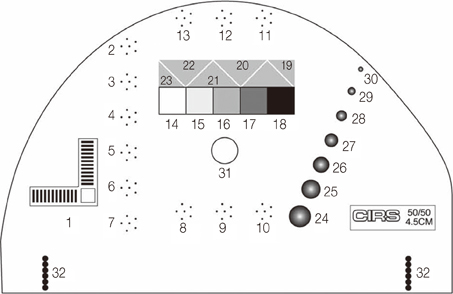
Figure 1 Schematic diagram of a Computerized Imaging Reference Systems (CIRS) phantom. The phantom breast detail component consists of 12 CaCO3 specks groups of different sizes, 7 hemispheric masses of different diameters, and 5 nylon fibers of different diameters.
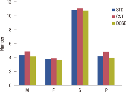
Figure 2 The image quality of all types among the three modes. Image quality is the number of fibers, specks, masses visualized, or image preference. STD=standard mode; CNT=contrast mode; DOSE=dose mode; M=mass; F=fiber; S=specks; P=image perference.
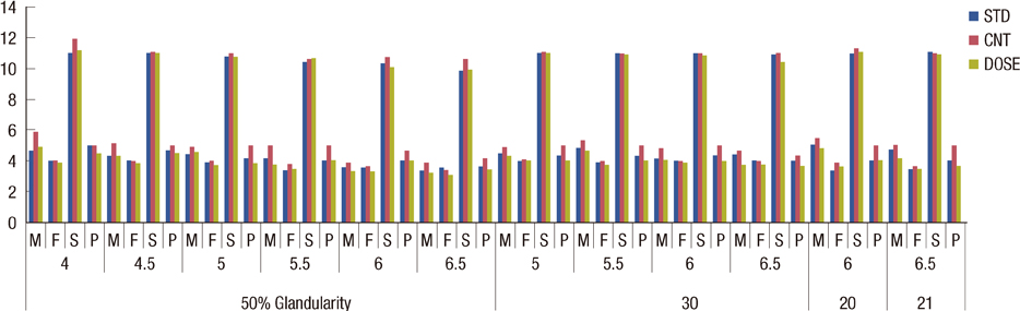
Figure 3 Relationship between image quality and breast thickness. STD=standard mode; CNT=contrast mode; DOSE=dose mode; M=mass; F=fiber; S=specks; P=image perference.
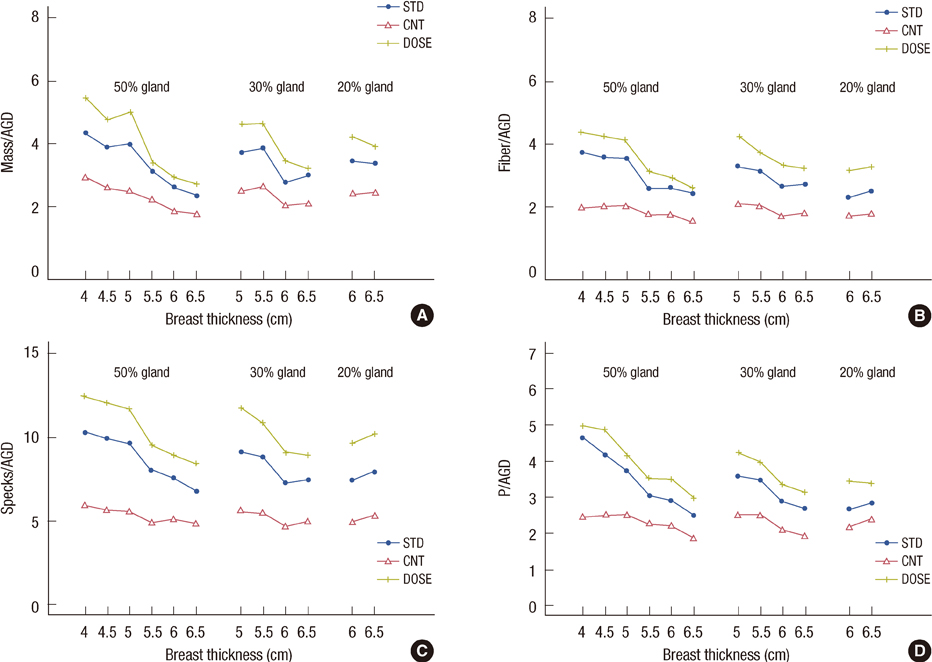
Figure 4 (A-D) The relationship between the image quality over a range of breast thickness and glandularity according to the automatic optimization of parameters (AOP) modes. Image quality is the dose-normalized scores of fibers, specks, masses visualized, or image preferences (lesion scores/average glandular dose [AGD]). STD=standard mode; CNT=contrast mode; DOSE=dose mode.
Reference
-
1. Jemal A, Siegel R, Ward E, Hao Y, Xu J, Thun MJ. Cancer statistics, 2009. CA Cancer J Clin. 2009. 59:225–249.
Article2. Vinnicombe S, Pinto Pereira SM, McCormack VA, Shiel S, Perry N, Dos Santos Silva IM. Full-field digital versus screen-film mammography: comparison within the UK breast screening program and systematic review of published data. Radiology. 2009. 251:347–358.
Article3. Ranger NT, Lo JY, Samei E. A technique optimization protocol and the potential for dose reduction in digital mammography. Med Phys. 2010. 37:962–969.
Article4. Samei E, Dobbins JT 3rd, Lo JY, Tornai MP. A framework for optimising the radiographic technique in digital X-ray imaging. Radiat Prot Dosimetry. 2005. 114:220–229.
Article5. Yaffe MJ, Mainprize JG. Detectors for digital mammography. Technol Cancer Res Treat. 2004. 3:309–324.
Article6. Huda W, Sajewicz AM, Ogden KM, Dance DR. Experimental investigation of the dose and image quality characteristics of a digital mammography imaging system. Med Phys. 2003. 30:442–448.
Article7. Chen B, Wang Y, Sun X, Guo W, Zhao M, Cui G, et al. Analysis of patient dose in full field digital mammography. Eur J Radiol. 2012. 81:868–872.
Article8. Williams MB, Raghunathan P, More MJ, Seibert JA, Kwan A, Lo JY, et al. Optimization of exposure parameters in full field digital mammography. Med Phys. 2008. 35:2414–2423.
Article9. Bor D, Tukel S, Olgar T, Aydin E. Variations in breast doses for an automatic mammography unit. Diagn Interv Radiol. 2008. 14:122–126.10. Berns EA, Hendrick RE, Cutter GR. Performance comparison of full-field digital mammography to screen-film mammography in clinical practice. Med Phys. 2002. 29:830–834.
Article11. Ministry of Health and Welfare. Recommendations of Radiation Dose to Patients from Mammography. 2008. Seoul: Ministry of Health and Welfare;1–46.12. Pachoud M, Lepori D, Valley JF, Verdun FR. A new test phantom with different breast tissue compositions for image quality assessment in conventional and digital mammography. Phys Med Biol. 2004. 49:5267–5281.
Article13. American College of Radiology, Committee on Quality Assurance in Mammography. Mammography Quality Control Manual: Radiologist's Section, Clinical Image Quality, Radiologic Technologist's Section, Medical Physicist's Section. 1999. Revised ed. Reston: American College of Radiology.14. Statement from the 1987 Como meeting of the International Commission on Radiological Protection. Radiology. 1988. 167:263–265.15. Ozdemir A. Clinical evaluation of breast dose and the factors affecting breast dose in screen-film mammography. Diagn Interv Radiol. 2007. 13:134–139.16. Pisano ED, Britt GG, Lin Y, Schell MJ, Burns CB, Brown ME. Factors affecting phantom scores at annual mammography facility inspections by the U.S. Food and Drug Administration. Acad Radiol. 2001. 8:864–870.
Article17. Obenauer S, Hermann KP, Grabbe E. Dose reduction in full-field digital mammography: an anthropomorphic breast phantom study. Br J Radiol. 2003. 76:478–482.
Article18. Gennaro G, Katz L, Souchay H, Alberelli C, di Maggio C. Are phantoms useful for predicting the potential of dose reduction in full-field digital mammography? Phys Med Biol. 2005. 50:1851–1870.
Article
- Full Text Links
-
- Actions
-
Cited
- CITED
-
- Close
- Share
-
- Similar articles
-
- Digital Mammography
- Which Phantom Is Better for Assessing the Image Quality in Full-Field Digital Mammography?: American College of Radiology Accreditation Phantom versus Digital Mammography Accreditation Phantom
- Comparison of Digital Mammography and Digital Breast Tomosynthesis
- Usefulness of a Small-Field Digital Mammographic Imaging System Using Parabolic Polycapillary Optics as a Diagnostic Imaging Tool: a Preliminary Study
- Mammography-Guided Interventional Procedure
