Which Phantom Is Better for Assessing the Image Quality in Full-Field Digital Mammography?: American College of Radiology Accreditation Phantom versus Digital Mammography Accreditation Phantom
- Affiliations
-
- 1Department of Radiology, Korea University College of Medicine, Ansan Hospital, Ansan 425-707, Korea. seoboky@korea.ac.kr
- 2Department of Radiology, Seoul National University College of Medicine, Seoul National University Hospital, Seoul 110-744, Korea.
- 3Department of Biostatistics, Korea University College of Medicine, Anam Hospital, Seoul 136-705, Korea.
- 4Department of Radiology, Korea University College of Medicine, Anam Hospital, Seoul 136-705, Korea.
- KMID: 1397509
- DOI: http://doi.org/10.3348/kjr.2012.13.6.776
Abstract
OBJECTIVE
To compare between the American College of Radiology (ACR) accreditation phantom and digital mammography accreditation phantom in assessing the image quality in full-field digital mammography (FFDM).
MATERIALS AND METHODS
In each week throughout the 42-week study, we obtained phantom images using both the ACR accreditation phantom and the digital mammography accreditation phantom, and a total of 42 pairs of images were included in this study. We assessed the signal-to-noise ratio (SNR) in each phantom image. A radiologist drew a square-shaped region of interest on the phantom and then the mean value of the SNR and the standard deviation were automatically provided on a monitor. SNR was calculated by an equation, measured mean value of SNR-constant coefficient of FFDM/standard deviation. Two breast radiologists scored visible objects (fibers, specks, and masses) with soft-copy images and calculated the visible rate (number of visible objects/total number of objects). We compared SNR and the visible rate of objects between the two phantoms and calculated the k-coefficient for interobserver agreement.
RESULTS
The SNR of the ACR accreditation phantom ranged from 42.0 to 52.9 (Mean, 47.3 +/- 2.79) and that of Digital Phantom ranged from 24.8 to 54.0 (Mean, 44.1 +/- 9.93) (p = 0.028). The visible rates of all three types of objects were much higher in the ACR accreditation phantom than those in the digital mammography accreditation phantom (p < 0.05). Interobserver agreement for visible rates of objects on phantom images was fair to moderate agreement (k-coefficients: 0.34-0.57).
CONCLUSION
The ACR accreditation phantom is superior to the digital mammography accreditation phantom in terms of SNR and visibility of phantom objects. Thus, ACR accreditation phantom appears to be satisfactory for assessing the image quality in FFDM.
Keyword
MeSH Terms
Figure
-
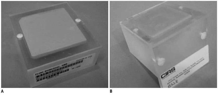
Fig. 1 Two mammography accreditation phantoms. Left is ACR Phantom (Nuclear Associates model 18-220) and right is Digital Phantom (CIRS Model 015DM). Digital Phantom has extended top edge allows ease of positioning on recumbent biopsy units. A. ACR Phantom. B. Digital Phanom. ACR = American College of Radiology

Fig. 2 Calculation of signal to noise ratio (SNR). Radiologist drew square-shaped region of interest (arrow) in background of phantom and then recorded mean value of SNR and standard deviation. SNR values were calculated based on following equation:
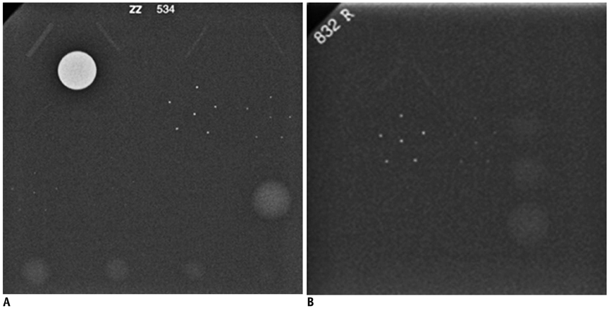
Fig. 3 SNR and visible rates on ACR and Digital Phantom images of week 23. A. On ACR Phantom, SNR is 45.23 and overall visible rate is 0.93 (15/16). Visible rates of fibers, specks, and masses are 1.00 (6/6), 0.80 (4/5), and 1.00 (5/5). B. On Digital Phantom, SNR is 32.47 and overall visible rate is 0.58 (7/12). Visible rates of fibers, specks, and masses are 0.50 (2/4), 0.50 (2/4), and 0.75 (3/4). Therefore, ACR Phantom is superior to Digital Phantom in terms of both SNR and visibility of phantom objects. SNR = signal to noise ratio, ACR = American College of Radiology
Reference
-
1. Feig SA, Yaffe MJ. Digital mammography. Radiographics. 1998. 18:893–901.2. Pisano ED, Gatsonis C, Hendrick E, Yaffe M, Baum JK, Acharyya S, et al. Diagnostic performance of digital versus film mammography for breast-cancer screening. N Engl J Med. 2005. 353:1773–1783.3. Pisano ED, Hendrick RE, Yaffe MJ, Baum JK, Acharyya S, Cormack JB, et al. Diagnostic accuracy of digital versus film mammography: exploratory analysis of selected population subgroups in DMIST. Radiology. 2008. 246:376–383.4. Fischmann A, Siegmann KC, Wersebe A, Claussen CD, Müller-Schimpfle M. Comparison of full-field digital mammography and film-screen mammography: image quality and lesion detection. Br J Radiol. 2005. 78:312–315.5. Krug KB, Stützer H, Girnus R, Zähringer M, Gossmann A, Winnekendonk G, et al. Image quality of digital direct flat-panel mammography versus an analog screen-film technique using a phantom model. AJR Am J Roentgenol. 2007. 188:399–407.6. American College of Radiology. Committee on Quality Assurance in Mammography. Mammography quality control manual: radiologist's section, clinical image quality, radiologic technologist's section, medical physicist's section. 1999. Rev. ed. Reston, VA: American College of Radiology.7. Butler PF. MQSA and accreditation for full-field digital mammography: everything you need to know in ½ hour. ACR Breast Imaging Accreditation Programs. 2012. Accessed September 28. Available at: http://www.acr.org/~/media/ACR/Documents/Accreditation/Mammography/MQSA%20and%20Accreditation%20RSNA07.pdf.8. McLean D, Eckert M, Heard R, Chan W. Review of the first 50 cases completed by the RACR mammography QA programme: phantom image quality, processor control and dose considerations. Australas Radiol. 1997. 41:387–391.9. Huda W, Sajewicz AM, Ogden KM, Scalzetti EM, Dance DR. How good is the ACR accreditation phantom for assessing image quality in digital mammography? Acad Radiol. 2002. 9:764–772.10. Jemal A, Clegg LX, Ward E, Ries LA, Wu X, Jamison PM, et al. Annual report to the nation on the status of cancer, 1975-2001, with a special feature regarding survival. Cancer. 2004. 101:3–27.11. Yamada T, Suzuki A, Uchiyama N, Ohuchi N, Takahashi S. Diagnostic performance of detecting breast cancer on computed radiographic (CR) mammograms: comparison of hard copy film, 3-megapixel liquid-crystal-display (LCD) monitor and 5-megapixel LCD monitor. Eur Radiol. 2008. 18:2363–2369.12. Kamitani T, Yabuuchi H, Matsuo Y, Setoguchi T, Sakai S, Okafuji T, et al. Diagnostic performance in differentiation of breast lesion on digital mammograms: comparison among hard-copy film, 3-megapixel LCD monitor, and 5-megapixel LCD monitor. Clin Imaging. 2011. 35:341–345.13. Chon KS, Park JG, Son HH, Kang SH, Park SH, Kim HW, et al. Usefulness of a small-field digital mammographic imaging system using parabolic polycapillary optics as a diagnostic imaging tool: a preliminary study. Korean J Radiol. 2009. 10:604–612.
- Full Text Links
-
- Actions
-
Cited
- CITED
-
- Close
- Share
-
- Similar articles
-
- Comparison Study of Image Quality of Direct and Indirect Conversion Digital Mammography System
- Review of Failed CT Phantom Image Evaluations in 2005 and 2006 by the CT Accreditation Program of the Korean Institute for Accreditation of Medical Image
- The Survey of Magnetic Resonance Imaging Quality according to Magnetic Field Strength in Korea
- Comparison of Image Quality between Mammography Dedicated Monitor and UHD 4K Monitor, Using Standard Mammographic Phantom: A Preliminary Study
- Dose Reduction in Automatic Optimization Parameter of Full Field Digital Mammography: Breast Phantom Study
