Surgical Anatomy of the Longus Colli Muscle and Uncinate Process in the Cervical Spine
- Affiliations
-
- 1Department of Orthopaedic Surgery, Hallym University Sacred Heart Hospital, Hallym University College of Medicine, Anyang, Korea. amhangpark@gmail.com
- 2Department of Orthopaedic Surgery, Yonsei University College of Medicine, Seoul, Korea.
- 3Department of Neurosurgery, Hallym University Sacred Heart Hospital, Hallym University College of Medicine, Anyang, Korea.
- 4Department of Orthopedic Surgery, Columbia University, The Spine Hospital at NY-Presbyterian/Allen Hospital, New York, NY, USA.
- KMID: 2374131
- DOI: http://doi.org/10.3349/ymj.2016.57.4.968
Abstract
- PURPOSE
There have been a few previous reports regarding the distances between the medial borders of the longus colli to expose the disc space. However, to our knowledge, there are no reports concerning longus colli dissection to expose the uncinate processes. This study was undertaken to assess the surgical relationship between the longus colli muscle and the uncinate process in the cervical spine.
MATERIALS AND METHODS
This study included 120 Korean patients randomly selected from 333 who had cervical spine MRIs and CTs from January 2003 to October 2013. They consisted of 60 males and 60 females. Each group was subdivided into six groups by age from 20 to 70 years or more. We measured three parameters on MRIs from C3 to T1: left and right longus colli distance and inter-longus colli distance. We also measured three parameters on CT: left and right uncinate distance and inter-uncinate distance.
RESULTS
The longus colli distances, uncinate distances, and inter-uncinate distances increased from C3 to T1. The inter-longus colli distances increased from C3 to C7. There was no difference in longus colli distances and uncinate distances between males and females. There was no difference in the six parameters for the different age groups.
CONCLUSION
Although approximate guidelines, we recommend the longus colli be dissected approximately 5 mm at C3-5, 6 mm at C5-6, 7 mm at C6-7, and 8 mm at C7-T1 to expose the uncinate process to its lateral edge.
Keyword
MeSH Terms
Figure
-
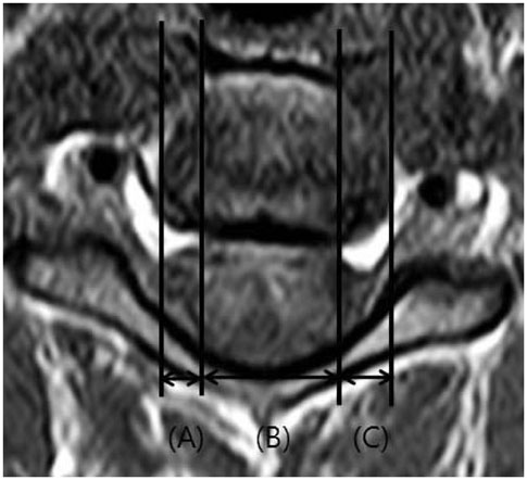
Fig. 1 Distance of longus colli muscle on axial image of MRI: (A) the distance from the lateral margin of the vertebral body to the medial margin of the right longus colli muscle (right longus colli distance), (B) the distance from the medial margin of the right longus colli muscle to that of the left longus colli muscle (inter-longus colli distance), and (C) the distance from the medial margin of the left longus colli muscle to the lateral margin of the vertebral body (left longus colli distance).
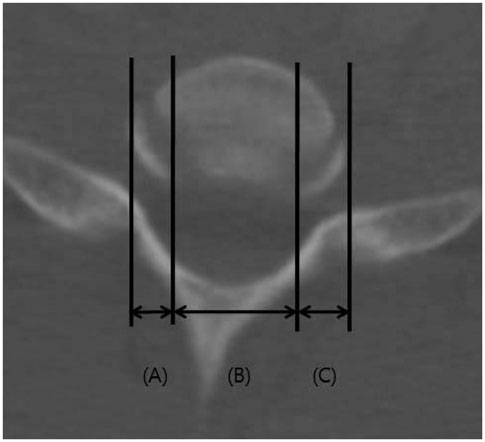
Fig. 2 Distance of uncinate processes on axial image of CT: (A) the distance from the lateral margin of the vertebral body to the medial margin of the right uncinate process (right uncinate distance), (B) the distance from the medial margin of the right uncinate process to that of the left uncinate process (inter-uncinate distance), and (C) the distance from the medial margin of the left uncinate process to the lateral margin of the vertebral body (left uncinate distance).
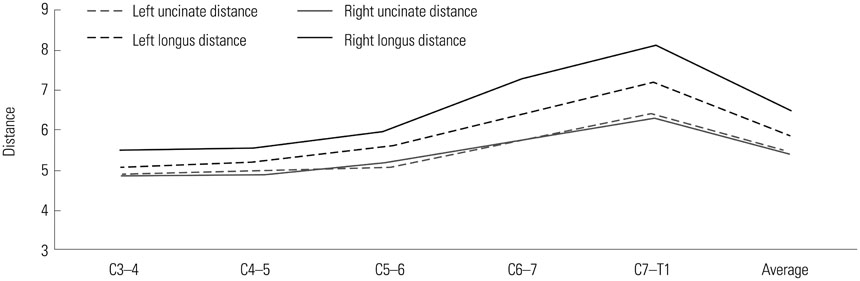
Fig. 3 Distance of longus colli muscles and uncinate processes according to cervical disc levels (mm).
Reference
-
1. Kiray A, Arman C, Naderi S, Güvencer M, Korman E. Surgical anatomy of the cervical sympathetic trunk. Clin Anat. 2005; 18:179–185.
Article2. Ebraheim NA, Lu J, Yang H, Heck BE, Yeasting RA. Vulnerability of the sympathetic trunk during the anterior approach to the lower cervical spine. Spine (Phila Pa 1976). 2000; 25:1603–1606.
Article3. Civelek E, Karasu A, Cansever T, Hepgul K, Kiris T, Sabanci A, et al. Surgical anatomy of the cervical sympathetic trunk during anterolateral approach to cervical spine. Eur Spine J. 2008; 17:991–995.
Article4. Kawashima M, Tanriover N, Rhoton AL Jr, Matsushima T. The transverse process, intertransverse space, and vertebral artery in anterior approaches to the lower cervical spine. J Neurosurg. 2003; 98:2 Suppl. 188–194.
Article5. Lu J, Ebraheim NA, Georgiadis GM, Yang H, Yeasting RA. Anatomic considerations of the vertebral artery: implications for anterior decompression of the cervical spine. J Spinal Disord. 1998; 11:233–236.6. Hong JT, Park DK, Lee MJ, Kim SW, An HS. Anatomical variations of the vertebral artery segment in the lower cervical spine: analysis by three-dimensional computed tomography angiography. Spine (Phila Pa 1976). 2008; 33:2422–2426.
Article7. Pait TG, Killefer JA, Arnautovic KI. Surgical anatomy of the anterior cervical spine: the disc space, vertebral artery, and associated bony structures. Neurosurgery. 1996; 39:769–776.
Article8. Javanshir K, Mohseni-Bandpei MA, Rezasoltani A, Amiri M, Rahgozar M. Ultrasonography of longus colli muscle: a reliability study on healthy subjects and patients with chronic neck pain. J Bodyw Mov Ther. 2011; 15:50–56.
Article9. Javanshir K, Rezasoltani A, Mohseni-Bandpei MA, Amiri M, Ortega-Santiago R, Fernández-de-Las-Peñas C. Ultrasound assessment of bilateral longus colli muscles in subjects with chronic bilateral neck pain. Am J Phys Med Rehabil. 2011; 90:293–301.
Article10. Lu J, Ebraheim NA, Yang H, Skie M, Yeasting RA. Cervical uncinate process: an anatomic study for anterior decompression of the cervical spine. Surg Radiol Anat. 1998; 20:249–252.
Article11. Uğur HC, Uz A, Attar A, Tekdemir I, Egemen N, Elhan A. Anatomical projection of the cervical uncinate process in ventral, ventrolateral, and posterior decompressive surgery. J Neurosurg. 2000; 93:2 Suppl. 248–251.
Article
- Full Text Links
-
- Actions
-
Cited
- CITED
-
- Close
- Share
-
- Similar articles
-
- Acute Calcific Tendinitis of the Longus Colli Muscle in the Cervical Spine
- Morphometry of the Uncinate Process, Vertebral Body, and Lamina of the C3–7 Vertebrae Relevant to Cervical Spine Surgery
- The Acute Calcific Prevertebral Tendinitis: Report of Two Cases
- Comparison of Ultrasonography and MRI in Measuring of Cervical Soft Tissue Structure
- Acute Calcific Prevertebral Tendinitis without Differentiated by Simple X-ray
