Korean J Gastroenterol.
2015 Mar;65(3):182-185. 10.4166/kjg.2015.65.3.182.
A Case of Pleomorphic Liposarcoma Originating from Mesentery
- Affiliations
-
- 1Department of Internal Medicine, Bundang Jesaeng General Hospital, Seongnam, Korea. parkjs@dmc.or.kr
- 2Department of Surgery, Bundang Jesaeng General Hospital, Seongnam, Korea.
- 3Department of Pathology, Bundang Jesaeng General Hospital, Seongnam, Korea.
- KMID: 2373205
- DOI: http://doi.org/10.4166/kjg.2015.65.3.182
Abstract
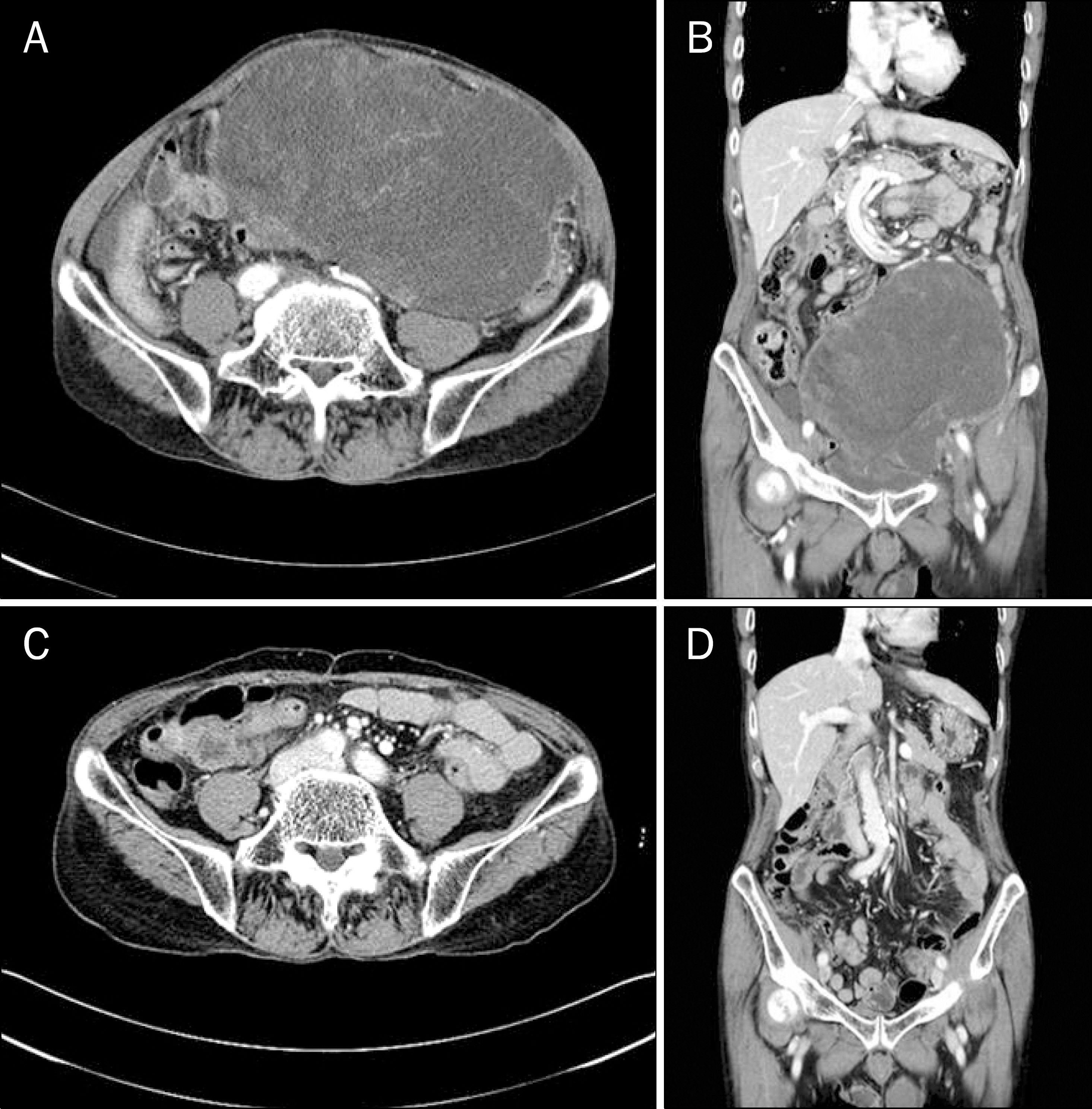
- Liposarcoma is one of the most common soft tissue sarcomas that occurs in adults and is currently divided into five main subgroups: well-differentiated, myxoid, round cell, pleomorphic, and dedifferentiated. Primary mesenteric liposarcoma is extremely rare, and the treatment strategy is surgical resection with a wide free margin, often followed by radiation and adjuvant chemotherapy if distant metastasis is not detected. A 73-year-old male patient presented with lower abdominal distension. Abdominal CT scan revealed a large homogeneously enhancing mass lesion abutting the sigmoid colon and urinary bladder. At laparotomy, the solid mass measured 28x26x12 cm in size, was well-demarcated, and originated from the mesentery of the middle ileum. It was removed along with some small intestine (ileocecal valve upper 50-150 cm) and ileal mesentery because of adhesion. Histologically, the tumor proved to be pleomorphic liposarcoma. The patient did not undergo any adjuvant treatment following surgery, but he remains disease free until 33 months after surgery. Herein, we report a case of pleomorphic liposarcoma arising from small bowel mesentery.
Keyword
MeSH Terms
Figure
Reference
-
References
1. Son BS, Seok SJ, Chung SK, et al. A case of liposarcoma arising in the mesentery. Korean J Gastroenterol. 2009; 54:243–247.
Article2. Cho YJ, Chun HJ, Park DK, et al. A case of dedifferentiated liposarcoma in retroperitoneum. Korean J Med. 2002; 62:552–556.3. Jain SK, Mitra A, Kaza RC, Malagi S. Primary mesenteric liposarcoma: An unusual presentation of a rare condition. J Gastrointest Oncol. 2012; 3:147–150.4. Grifasi C, Calogero A, Carlomagno N, Campione S, D'Armiento FP, Renda A. Intraperitoneal dedifferentiated liposarcoma showing MDM2 amplification: case report. World J Surg Oncol. 2013; 11:305.
Article5. Shen Z, Wang S, Fu L, et al. Therapeutic experience with primary liposarcoma from the sigmoid mesocolon accompanied with well-differentiated liposarcomas in the pelvis. Surg Today. 2014; 44:1863–1868.
Article6. Liu Y, Ishibashi H, Sako S, et al. A giant mesentery malignant soli-tary fibrous tumor recurring as dedifferentiated liposarcoma- a report of a very rare case and literature review. Gan To Kagaku Ryoho. 2013; 40:2466–2469.7. Eltweri AM, Gravante G, Read-Jones SL, Rai S, Bowrey DJ, Haynes IG. A case of recurrent mesocolon myxoid liposarcoma and review of the literature. Case Rep Oncol Med. 2013; 2013:692754.
Article8. Khan MI, Zafar A, Younas M, Malik I. Huge mesenteric liposarcoma. J Pak Med Assoc. 2013; 63:775–777.9. Garg PK, Jain BK, Dahiya D, Bhatt S, Arora VK. Mesenteric liposarcoma: report of two cases with review of literature. J Gastrointest Cancer. 2014; 45(Suppl 1):170–174.
Article10. Gebhard S, Coindre JM, Michels JJ, et al. Pleomorphic liposarcoma: clinicopathologic, immunohistochemical, and fol-low-up analysis of 63 cases: a study from the French Federation of Cancer Centers Sarcoma Group. Am J Surg Pathol. 2002; 26:601–616.11. Dalal KM, Kattan MW, Antonescu CR, Brennan MF, Singer S. Subtype specific prognostic nomogram for patients with primary liposarcoma of the retroperitoneum, extremity, or trunk. Ann Surg. 2006; 244:381–391.
Article12. Dubin MR, Chang EW. Liposarcoma of the tongue: case report and review of the literature. Head Face Med. 2006; 2:21.
Article13. Choi CW, Kim ST, Kim HJ, et al. Myxoid Liposarcoma of the Parietal Peritoneum. Korean J Gastroenterol. 1999; 33:298–302.14. Ishiguro S, Yamamoto S, Chuman H, Moriya Y. A case of resected huge ileocolonic mesenteric liposarcoma which responded to pre-operative chemotherapy using doxorubicin, cisplatin and ifosfamide. Jpn J Clin Oncol. 2006; 36:735–738.
Article15. Nakamura A, Tanaka S, Takayama H, et al. A mesenteric liposarcoma with production of granulocyte colony-stimulating factor. Intern Med. 1998; 37:884–890.
Article


