Korean J Gastroenterol.
2015 Feb;65(2):127-131. 10.4166/kjg.2015.65.2.127.
Appendiceal Neuroendocrine Tumor with Lymph Node Metastasis in a Teenager
- Affiliations
-
- 1Department of Surgery, Wonkwang University School of Medicine, Iksan, Korea. parkwc@wonkwang.ac.kr
- KMID: 2373195
- DOI: http://doi.org/10.4166/kjg.2015.65.2.127
Abstract
- Neuroendocrine tumor (NET) is a cancer-like tumor that occurs mostly in the gastrointestinal system. Within the gastrointestinal tract, NET most commonly occurs in the rectum whereas appendix is very rarely involved. In most cases of appendiceal NET, it is found at a relatively early stage compared to other NETs because appendiceal NET frequently presents with acute appendicitis because appendiceal NET frequently presents with acute appendicitis even when the size is smaller than 1 cm. Therefore, it is very rare for lymph node metastasis to occur in a young adult. Herein, we report a rare case of grade 1 appendiceal NET with lymph node metastasis which developed in a teenage male.
MeSH Terms
Figure
-

Fig. 1. CT shows dilated appendix, which measures up to 2 cm in diameter (arrow), with proximal appendicolith.
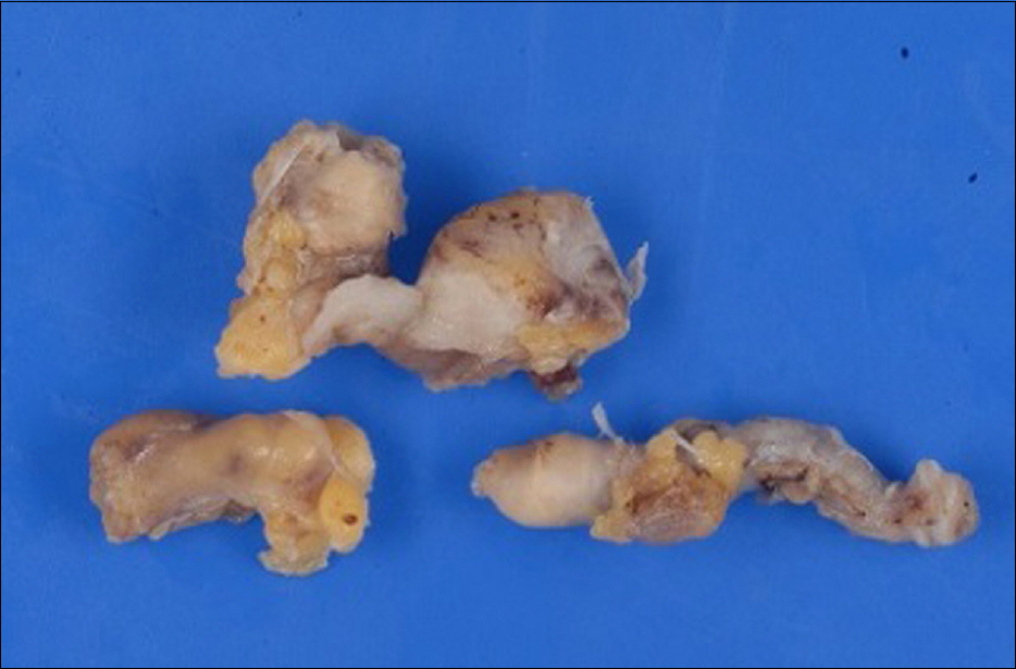
Fig. 2. Gross findings of the resected appendix. No definite tumor is observed on the specimen.
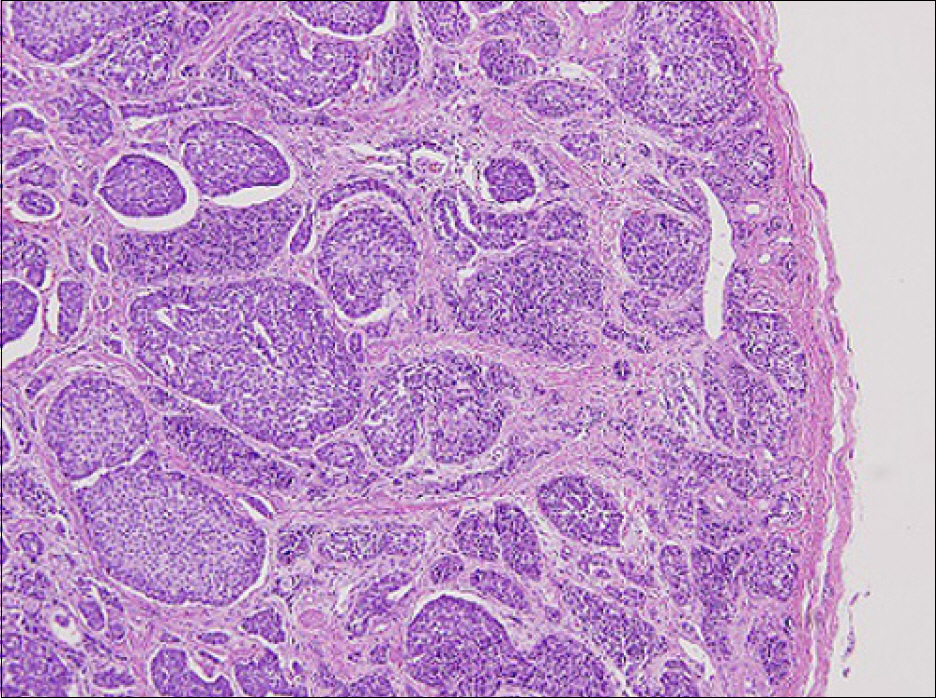
Fig. 3. Microscopic findings. On histologic examination, the appendiceal lesion is a Grade 1 neuroendocrine tumor with no lymphova-scular invasion or cellular atypism. Mitotic count is lower than 2/10 HPF (H&E, ×100).
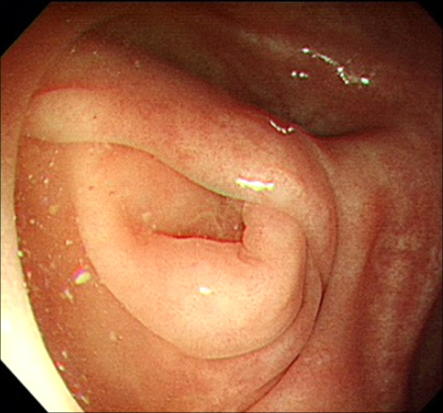
Fig. 4. Colonoscopy finding shows a normal appearing appendiceal orifice except for mild mucosal inflammation.
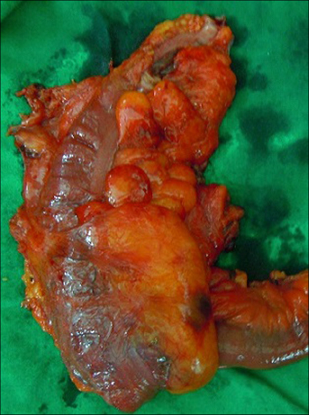
Fig. 5. Gross specimen obtained after right hemicolectomy.
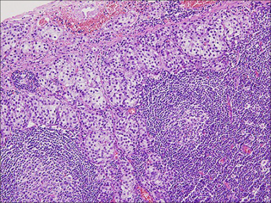
Fig. 6. Microcopic finding of lymph node (H&E,×200).
Reference
-
References
1. Kang BS, Kim JW. Gastrointestinal carcinoid tumor: clinical review of 36 cases. J Korean Surg Soc. 2009; 76:1–6.
Article2. Lee MH, Shin SJ, Jeon SJ, et al. Clinical characteristics of gastrointestinal carcinoid tumors. Korean J Gastrointest Endosc. 2010; 40:347–351.3. Eggenberger JC. Carcinoid and other neuroendocrine tumors of the colon and rectum. Clin Colon Rectal Surg. 2011; 24:129–134.
Article4. Heo SC, Ahn YJ, Jung IM, et al. Appendiceal carcinoids detected in patients with symptoms of acute appendicitis. J Korean Surg Soc. 2004; 67:146–151.5. Song SK, Choi ST, Kim KK, et al. Clinical review of appendiceal tumors (retrospective study of 3,744 appendectomies or right hemicolectomies). J Korean Surg Soc. 2007; 73:42–47.6. Shin HD. Diagnosis and treatment of rectal neuroendocrine tumor. Korean J Med. 2014; 87:415–423.
Article7. Kim GU, Ye BD, Byeon JS, et al. Endoscopic resection for rectal carcinoid tumor: efficacy and clinical results of follow-up. Intest Res. 2011; 9:217–224.
Article8. Park CH, Cheon JH, Kim JO, et al. Criteria for decision making after endoscopic resection of well-differentiated rectal carcinoids with regard to potential lymphatic spread. Endoscopy. 2011; 43:790–795.
Article9. Lee DH, Chin HM, Kim JG, Lee YB, Park WB, Chun CS. Clinical analysis of carcinoid tumors. J Korean Surg Soc. 1997; 53:315–323.10. Chang NS, Sung KC, Pyeon YJ, et al. Clinical reviews of patients with carcinoid tumor. Korean J Gastroenterol. 1997; 30:179–186.
- Full Text Links
-
- Actions
-
Cited
- CITED
-
- Close
- Share
-
- Similar articles
-
- A small, low-grade rectal neuroendocrine tumor with lateral pelvic lymph node metastasis: a case report
- Metastatic Large Cell Neuroendocrine Carcinoma Combined with Gastric Adenocarcinoma
- MRI Findings of an Ampulla of Vater Neuroendocrine Tumor with Liver and Lymph Node Metastasis: a Case Report
- Natural Course of an Untreated Metastatic Perirectal Lymph Node After the Endoscopic Resection of a Rectal Neuroendocrine Tumor
- Pattern of Cervical Neck Lymph Node Metastasis in Papillary Thyroid Carcinoma according to Tumor Size
