Improved accuracy in periodontal pocket depth measurement using optical coherence tomography
- Affiliations
-
- 1Department of Periodontology, Seoul National University School of Dentistry, Seoul, Korea. periopf@snu.ac.kr
- 2Department of Biomedical Radiation Sciences, Seoul National University Graduate School of Convergence Science and Technology, Seoul, Korea.
- 3Department of Health Policy and Management, Korea University College of Health Sciences, Seoul, Korea.
- 4Dental Research Institute, Seoul National University School of Dentistry, Seoul, Korea.
- 5Department of Oral and Maxillofacial Radiology, Seoul National University School of Dentistry, Seoul, Korea.
- KMID: 2368883
- DOI: http://doi.org/10.5051/jpis.2017.47.1.13
Abstract
- PURPOSE
The purpose of this study was to examine whether periodontal pocket could be satisfactorily visualized by optical coherence tomography (OCT) and to suggest quantitative methods for measuring periodontal pocket depth.
METHODS
We acquired OCT images of periodontal pockets in a porcine model and determined the actual axial resolution for measuring the exact periodontal pocket depth using a calibration method. Quantitative measurements of periodontal pockets were performed by real axial resolution and compared with the results from manual periodontal probing.
RESULTS
The average periodontal pocket depth measured by OCT was 3.10±0.15 mm, 4.11±0.17 mm, 5.09±0.17 mm, and 6.05±0.21 mm for each periodontal pocket model, respectively. These values were similar to those obtained by manual periodontal probing.
CONCLUSIONS
OCT was able to visualize periodontal pockets and show attachment loss. By calculating the calibration factor to determine the accurate axial resolution, quantitative standards for measuring periodontal pocket depth can be established regardless of the position of periodontal pocket in the OCT image.
Keyword
MeSH Terms
Figure
-

Figure 1 The porcine mandibular sample positioned for OCT imaging acquisition (A) and the OCT images obtained of the target area (B) showing the dental structures. OCT, optical coherence tomography; G, gingiva; E, enamel, D, dentin; PP, periodontal pocket.

Figure 2 The porcine sample for calibration of the axial resolution of the OCT image and the calibration phantom between the gingiva and the tooth surface (A). OCT images of the sample with 0° (B) and 20° (C) of imaging inclination. OCT, optical coherence tomography
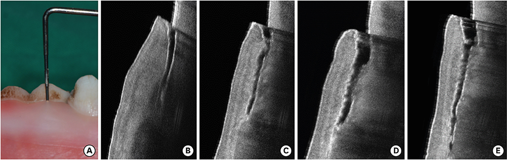
Figure 3 Measuring the periodontal pocket depth of a porcine sample by manual probing (A) and the OCT images of each sample at the depths of 3 mm (B), 4 mm (C), 5 mm (D), and 6 mm (E). OCT, optical coherence tomography.
Cited by 1 articles
-
Quantitative measurement of peri-implant bone defects using optical coherence tomography
Sulhee Kim, Se-Ryong Kang, Hee-Jung Park, Bome Kim, Tae-Il Kim, Won-Jin Yi
J Periodontal Implant Sci. 2018;48(2):84-91. doi: 10.5051/jpis.2018.48.2.84.
Reference
-
1. Mota CC, Fernandes LO, Cimões R, Gomes AS. Non-invasive periodontal probing through fourier-domain optical coherence tomography. J Periodontol. 2015; 86:1087–1094.
Article2. Huang D, Swanson EA, Lin CP, Schuman JS, Stinson WG, Chang W, et al. Optical coherence tomography. Science. 1991; 254:1178–1181.
Article3. Adhi M, Duker JS. Optical coherence tomography--current and future applications. Curr Opin Ophthalmol. 2013; 24:213–221.4. Sattler E, Kästle R, Welzel J. Optical coherence tomography in dermatology. J Biomed Opt. 2013; 18:061224.
Article5. Kirtane TS, Wagh MS. Endoscopic optical coherence tomography (OCT): advances in gastrointestinal imaging. Gastroenterol Res Pract. 2014; 2014:376367.
Article6. Ferrante G, Presbitero P, Whitbourn R, Barlis P. Current applications of optical coherence tomography for coronary intervention. Int J Cardiol. 2013; 165:7–16.
Article7. Cheng HM, Guitera P. Systematic review of optical coherence tomography usage in the diagnosis and management of basal cell carcinoma. Br J Dermatol. 2015; 173:1371–1380.
Article8. Colston BW Jr, Everett MJ, Da Silva LB, Otis LL, Stroeve P, Nathel H. Imaging of hard- and soft-tissue structure in the oral cavity by optical coherence tomography. Appl Opt. 1998; 37:3582–3585.
Article9. Feldchtein F, Gelikonov V, Iksanov R, Gelikonov G, Kuranov R, Sergeev A, et al. In vivo OCT imaging of hard and soft tissue of the oral cavity. Opt Express. 1998; 3:239–250.10. Imai K, Shimada Y, Sadr A, Sumi Y, Tagami J. Noninvasive cross-sectional visualization of enamel cracks by optical coherence tomography in vitro . J Endod. 2012; 38:1269–1274.
Article11. Ishibashi K, Ozawa N, Tagami J, Sumi Y. Swept-source optical coherence tomography as a new tool to evaluate defects of resin-based composite restorations. J Dent. 2011; 39:543–548.
Article12. Shimada Y, Sadr A, Burrow MF, Tagami J, Ozawa N, Sumi Y. Validation of swept-source optical coherence tomography (SS-OCT) for the diagnosis of occlusal caries. J Dent. 2010; 38:655–665.
Article13. Di Stasio D, Lauritano D, Romano A, Salerno C, Minervini G, Minervini G, et al. In vivo characterization of oral pemphigus vulgaris by optical coherence tomography. J Biol Regul Homeost Agents. 2015; 29:39–41.14. Tsai MT, Lee CK, Lee HC, Chen HM, Chiang CP, Wang YM, et al. Differentiating oral lesions in different carcinogenesis stages with optical coherence tomography. J Biomed Opt. 2009; 14:044028.
Article15. Hsieh YS, Ho YC, Lee SY, Lu CW, Jiang CP, Chuang CC, et al. Subgingival calculus imaging based on swept-source optical coherence tomography. J Biomed Opt. 2011; 16:071409.
Article16. Kao MC, Lin CL, Kung CY, Huang YF, Kuo WC. Miniature endoscopic optical coherence tomography for calculus detection. Appl Opt. 2015; 54:7419–7423.
Article17. Baek JH, Na J, Lee BH, Choi E, Son WS. Optical approach to the periodontal ligament under orthodontic tooth movement: a preliminary study with optical coherence tomography. Am J Orthod Dentofacial Orthop. 2009; 135:252–259.
Article18. Fernandes LO, Mota CC, de Melo LS, da Costa Soares MU, da Silva Feitosa D, Gomes AS. In vivo assessment of periodontal structures and measurement of gingival sulcus with optical coherence tomography: a pilot study. J Biophotonics. Forthcoming. 2016.19. Agrawal P, Sanikop S, Patil S. New developments in tools for periodontal diagnosis. Int Dent J. 2012; 62:57–64.
Article20. Fujimoto JG. Optical coherence tomography for ultrahigh resolution in vivo imaging. Nat Biotechnol. 2003; 21:1361–1367.
Article21. Armitage GC. Clinical evaluation of periodontal diseases. Periodontol 2000. 1995; 7:39–53.
Article22. Savage A, Eaton KA, Moles DR, Needleman I. A systematic review of definitions of periodontitis and methods that have been used to identify this disease. J Clin Periodontol. 2009; 36:458–467.
Article23. Xiang X, Sowa MG, Iacopino AM, Maev RG, Hewko MD, Man A, et al. An update on novel non-invasive approaches for periodontal diagnosis. J Periodontol. 2010; 81:186–198.
Article24. Kao RT, Pasquinelli K. Thick vs. thin gingival tissue: a key determinant in tissue response to disease and restorative treatment. J Calif Dent Assoc. 2002; 30:521–526.25. Thoma DS, Mühlemann S, Jung RE. Critical soft-tissue dimensions with dental implants and treatment concepts. Periodontol 2000. 2014; 66:106–118.
Article26. Lee A, Fu JH, Wang HL. Soft tissue biotype affects implant success. Implant Dent. 2011; 20:e38–47.
Article27. Grossi SG, Dunford RG, Ho A, Koch G, Machtei EE, Genco RJ. Sources of error for periodontal probing measurements. J Periodontal Res. 1996; 31:330–336.
Article28. van der Velden U, de Vries JH. The influence of probing force on the reproducibility of pocket depth measurements. J Clin Periodontol. 1980; 7:414–420.
Article29. Badersten A, Nilvéus R, Egelberg J. Reproducibility of probing attachment level measurements. J Clin Periodontol. 1984; 11:475–485.
Article30. Watts TL. Probing site configuration in patients with untreated periodontitis. A study of horizontal positional error. J Clin Periodontol. 1989; 16:529–533.
Article31. Hsieh YS, Ho YC, Lee SY, Chuang CC, Tsai JC, Lin KF, et al. Dental optical coherence tomography. Sensors (Basel). 2013; 13:8928–8949.
Article32. Goodson JM, Haffajee AD, Socransky SS. The relationship between attachment level loss and alveolar bone loss. J Clin Periodontol. 1984; 11:348–359.
Article
- Full Text Links
-
- Actions
-
Cited
- CITED
-
- Close
- Share
-
- Similar articles
-
- Comparisons of the diagnostic accuracies of optical coherence tomography, micro-computed tomography, and histology in periodontal disease: an ex vivo study
- Quantitative measurement of peri-implant bone defects using optical coherence tomography
- Availability of Optical Coherence Tomography in Diagnosis and Classification of Choroidal Neovascularization
- Choroidal Thickness at the Outside of Fovea in Diabetic Retinopathy Using Spectral-Domain Optical Coherence Tomography
- Optical Imaging and Its Clinical Application in Otorhinolaryngology
