Ann Dermatol.
2016 Dec;28(6):787-788. 10.5021/ad.2016.28.6.787.
Pseudoaneurysm as a Post-Biopsy Complication
- Affiliations
-
- 1Department of Dermatology, Korea University Guro Hospital, Seoul, Korea. drsshong@hanmail.net
- 2Department of Radiology, Korea University Guro Hospital, Seoul, Korea.
- KMID: 2368140
- DOI: http://doi.org/10.5021/ad.2016.28.6.787
Abstract
- No abstract available.
MeSH Terms
Figure
-
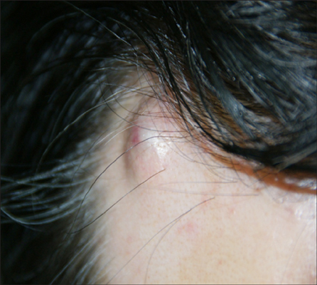
Fig. 1 A 1×1.1-cm-sized pulsating mass with intermittent pain.
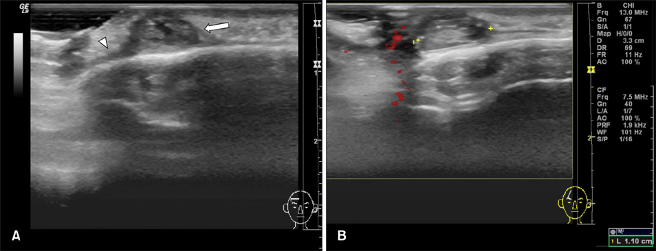
Fig. 2 Mixed echoic subcutaneous lumen (arrow) with fistula (arrowhead) shown on left side. (A) Transverse view, (B) longitudinal view.
Reference
-
1. Cook JL, Perone JB. A prospective evaluation of the incidence of complications associated with Mohs micrographic surgery. Arch Dermatol. 2003; 139:143–152.
Article2. Ito T, Nakahara T, Takeuchi S, Uchi H, Takahara M, Moroi Y, et al. Four cases of successfully treated chronic expanding soft tissue hematoma. Ann Dermatol. 2014; 26:107–110.
Article3. Bordeaux JS, Martires KJ, Goldberg D, Pattee SF, Fu P, Maloney ME. Prospective evaluation of dermatologic surgery complications including patients on multiple antiplatelet and anticoagulant medications. J Am Acad Dermatol. 2011; 65:576–583.
Article4. Rubio-Palau J, Ferrer-Fuertes A, García-Díez E, Garcia-Linares J, Martí-Pagès C, Sieira-Gil R. Traumatic pseudoaneurysm of the superficial temporal artery: case report and review of the literature. Oral Surg Oral Med Oral Pathol Oral Radiol. 2014; 117:e112–e114.
Article5. Dunbar SW, Hurst EA. Pseudoaneurysm formation and repair after Mohs micrographic surgery: a report of 3 cases. JAMA Dermatol. 2014; 150:546–549.
Article
- Full Text Links
-
- Actions
-
Cited
- CITED
-
- Close
- Share
-
- Similar articles
-
- Pseudoaneurysm of the Breast after 14-Gauge Core Biopsy: Case Report
- Pseudoaneurysm of the Breast after Core Needle Biopsy: A Case Report
- A Case of Pseudoaneurysm of the Superior Thyroid Artery after Core Needle Biopsy
- Spontaneous Resolution of Pulmonary Artery Pseudoaneurysm after Tube Thoracostomy
- Gastroduodenal artery pseudoaneurysm in chronic pancreatitis: diagnosis with duplex US and CT: a case report
