Morphologic Changes of Zebrafish Melanophore after Intense Pulsed Light and Q-Switched Nd:YAG Laser Irradiation
- Affiliations
-
- 1Department of Dermatology, Korea University College of Medicine, Seoul, Korea. kumcihk@korea.ac.kr
- 2Laboratory of Neurodevelopmental Genetics, Graduate School of Medicine, Korea University, Seoul, Korea.
- 3Department of Anatomy, Korea University College of Medicine, Seoul, Korea.
- KMID: 2368121
- DOI: http://doi.org/10.5021/ad.2016.28.6.711
Abstract
- BACKGROUND
Recently, the pulse-in-pulse mode of intense pulsed light (IPL) has been used increasingly for the treatment of melasma.
OBJECTIVE
To observe the morphologic changes in the melanophore in adult zebrafish after irradiation with conventional and pulse-in-pulse IPL and Q-switched Nd:YAG (QSNY) laser.
METHODS
Adult zebrafish were irradiated with conventional and pulse-in-pulse mode of IPL. The conditions for conventional IPL were 3 mJ/cm², 560 nm filter, and pulse widths of 7, 20, and 35 msec. The pulse-in-pulse conditions were 3 mJ/cm² and on-time 1/off-time 2. The QSNY laser was used with the settings of 1,064 nm, 0.4 J/cm², a 7 mm spot size, and one shot. Specimens were observed using a light microscope, a transmission electron microscope (TEM), a scanning electron microscope (SEM) and a confocal microscope.
RESULTS
After conventional IPL irradiation with a 7 msec pulse width, melanophore breakage was observed using light microscopy. Under TEM, irradiation with conventional IPL for 7 msec and pulse-in-pulse IPL induced melanophore thermolysis with vacuolization. However, changes in the melanophore were not observed with 35 msec IPL. Under SEM, unlike the control and QSNY groups, IPL-irradiated zebrafish showed finger-like fusion in the protein structure of scales. Specimens examined by a confocal microscope after conventional IPL irradiation showed a larger green-stained area on TUNEL staining than that after pulse-in-pulse mode IPL irradiation.
CONCLUSION
Zebrafish irradiated with long pulse-IPL showed no morphologic changes using light microscopy, while morphological changes in melanophores were evident with use of TEM. Pulse-in-pulse mode IPL caused less damage than conventional IPL.
Keyword
MeSH Terms
Figure
-
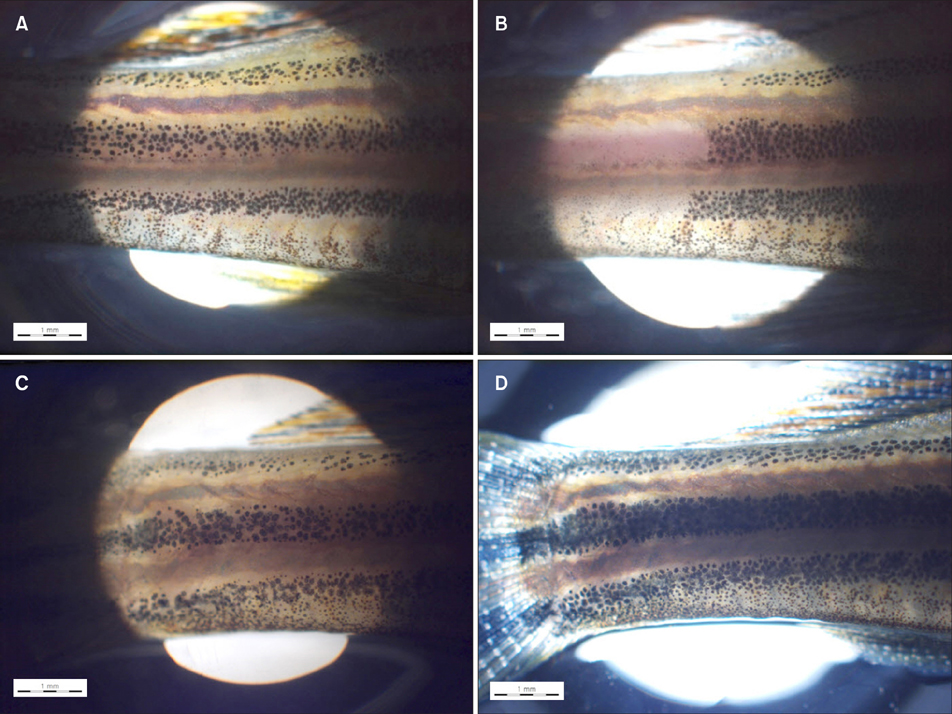
Fig. 1 Light microscopy in zebrafish before (A) and 1 week after (B) intense pulsed light (IPL) irradiation with 560 nm filter and 7 msec, and before (C) and 1 week after (D) IPL irradiation with 560 nm filter and 35 msec. Conventional IPL irradiation with a 7 msec pulse width induced melanophore breakage. However, the clearance of melanophore was not observed after irradiation with a 35 msec pulse width.

Fig. 2 Representative transmission electron microscope images of (A) control, (B) pulse-in-pulse mode intense pulsed light (IPL), (C) IPL irradiation with 560 nm filter and 7 msec, and (D) IPL irradiation with 560 nm filter and 35 msec. (B, C) Irradiation with pulse-in-pulse IPL and conventional IPL for 7 msec induced melanophore thermolysis (black arrowhead), with vacuolization (white arrowhead), central electron lucency (white arrow), and empty spaces (black arrow) evident due to melanophore destruction. Conventional IPL irradiation with 35 msec pulse width caused some melanophore changes but no vacuolization. Scale bar=100 µm.
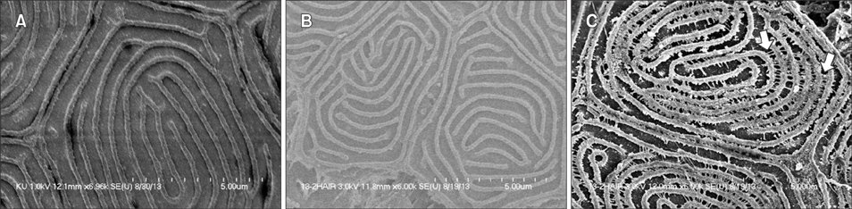
Fig. 3 Representative scanning electron microscope images of (A) control, (B) Q-switched Nd:YAG (QSNY) laser irradiation, and (C) intense pulsed light (IPL) irradiation with 560 nm filter and 20 msec. Control zebrafish or zebrafish irradiated using a QSNY laser did not show microstructural changes. However, IPL-irradiated zebrafish showed finger-like fusion (arrows) in the protein structure of scales.

Fig. 4 Representative confocal microscopic findings of (A) control, (B) pulse-in-pulsed mode intense pulsed light (IPL), and (C) conventional mode IPL irradiation with 560 nm filter and 7 msec. After conventional IPL irradiation, zebrafish showed a larger green-stained area on TUNEL staining than that after pulse-in-pulse mode IPL irradiation (×20).
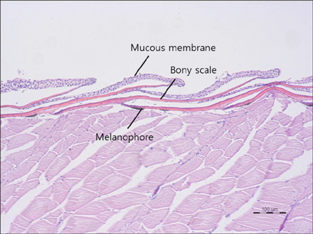
Fig. 5 Normal adult zebrafish skin stained with H&E stain at ×100 magnification. Unlike the human skin, zebrafish have scales which are calcified plates originating in the dermis and covered by the mucous membrane. A horizontal view of fish skin reveals melanophores right below the scales.
Reference
-
1. Chung JY, Choi M, Lee JH, Cho S, Lee JH. Pulse in pulse intense pulsed light for melasma treatment: a pilot study. Dermatol Surg. 2014; 40:162–168.
Article2. Ni-Komatsu L, Orlow SJ. Identification of novel pigmentation modulators by chemical genetic screening. J Invest Dermatol. 2007; 127:1585–1592.
Article3. Li Q, Uitto J. Zebrafish as a model system to study skin biology and pathology. J Invest Dermatol. 2014; 134:e21.
Article4. Na SY, Cho S, Lee JH. Intense pulsed light and low-fluence Q-switched Nd:YAG laser treatment in Melasma patients. Ann Dermatol. 2012; 24:267–273.
Article5. Yamashita T, Negishi K, Hariya T, Kunizawa N, Ikuta K, Yanai M, et al. Intense pulsed light therapy for superficial pigmented lesions evaluated by reflectance-mode confocal microscopy and optical coherence tomography. J Invest Dermatol. 2006; 126:2281–2286.
Article6. Anderson RR, Margolis RJ, Watenabe S, Flotte T, Hruza GJ, Dover JS. Selective photothermolysis of cutaneous pigmentation by Q-switched Nd: YAG laser pulses at 1064, 532, and 355 nm. J Invest Dermatol. 1989; 93:28–32.
Article7. Wenzel S, Landthaler M, Baumler W. Recurring mistakes in tattoo removal. A case series. Dermatology. 2009; 218:164–167.8. Altshuler GB, Anderson RR, Manstein D, Zenzie HH, Smirnov MZ. Extended theory of selective photothermolysis. Lasers Surg Med. 2001; 29:416–432.
Article9. Negishi K, Kushikata N, Tezuka Y, Takeuchi K, Miyamoto E, Wakamatsu S. Study of the incidence and nature of "very subtle epidermal melasma" in relation to intense pulsed light treatment. Dermatol Surg. 2004; 30:881–886. discussion 886.
Article10. Yun WJ, Moon HR, Lee MW, Choi JH, Chang SE. Combination treatment of low-fluence 1,064-nm Q-switched Nd: YAG laser with novel intense pulse light in Korean melasma patients: a prospective, randomized, controlled trial. Dermatol Surg. 2014; 40:842–850.11. Park JH, Kim JI, Kim WS. Treatment of persistent facial postinflammatory hyperpigmentation with novel pulse-inpulse mode intense pulsed light. Dermatol Surg. 2016; 42:218–224.
Article12. Kang HY. Melasma and aspects of pigmentary disorders in Asians. Ann Dermatol Venereol. 2012; 139:Suppl 4. S144–S147.
Article13. Kim MJ, Kim JS, Cho SB. Punctate leucoderma after melasma treatment using 1064-nm Q-switched Nd:YAG laser with low pulse energy. J Eur Acad Dermatol Venereol. 2009; 23:960–962.
Article
- Full Text Links
-
- Actions
-
Cited
- CITED
-
- Close
- Share
-
- Similar articles
-
- Treatment of a Congenital Melanocytic Nevus by New Combined Therapy: Intense Pulsed Light and a Q-switched Nd:YAG Laser
- Intense Pulsed Light and Q-Switched 1,064-nm Neodymium-Doped Yttrium Aluminum Garnet Laser Treatment for the Scarring Lesion of Discoid Lupus Erythematosus
- Split-face Comparison of Pulse-in-pulse Type Intense Pulsed Light Versus Low-fluence Multi-pass 1064 nm Q-switched Nd:YAG Laser in the Treatment of Facial Melasma
- Treatment of acne fulminans with intense pulsed light: a case report
- Intense Pulsed Light and Low-Fluence Q-Switched Nd:YAG Laser Treatment in Melasma Patients
