Refractory Spontaneous Chronic Subdural Hematoma: A Rare Presentation of an Intracranial Arteriovenous Fistula
- Affiliations
-
- 1Department of Neurosurgery, Dongsan Medical Center, Keimyung University School of Medicine, Daegu, Korea. bach1158@dsmc.or.kr
- KMID: 2367329
- DOI: http://doi.org/10.7461/jcen.2016.18.4.373
Abstract
- The author has encountered a 67-year-old man with dural arteriovenous fistula (AVF) presenting as a non-traumatic chronic subdural hematoma (CSDH). This previously healthy patient was hospitalized due to progressive headache with subacute onset. He underwent burr-hole surgery twice for evacuating the left CSDH that was thickest at the posterior temporal area. The operative procedure and finding was not extraordinary, but subdural hematoma slowly progressed for days following the revision surgery. After investigation by super-selective external carotid angiography, a dural AVF found near the transverse-sigmoid sinus was diagnosed. Dural AVF was completely occluded with trans-arterial injecting polyvinyl alchol particles into the petrosquamosal branch of the middle meningeal artery. The patient showed a good neurological outcome with no additional intervention. Brain surgeons have to consider the possibility of dural AVF and perform cerebral angiogram if necessary when they manage the cases that have a spontaneously occurred and repeatedly recurring CSDH.
Keyword
MeSH Terms
Figure
-
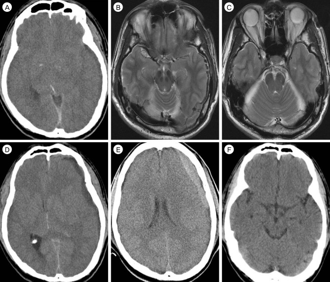
Fig. 1 (A) Unenhanced computed tomography (CT) taken at the initial presentation shows a chronic subdural hematoma in the left hemisphere with midline displacement. (B, C) Magnetic resonance images reveal no subarachnoid bleeding and normal transverse-sigmoid sinuses. (D) Repeated CT on the second admission demonstrates recurrence of an isoodense left subdural hematoma. (E) Recollection of a new hematoma is occurred 7 days later following the revision burr-hole surgery. (F) Brain CT scan obtained after treatments depicts complete resolution of the subdural hemorrhage.
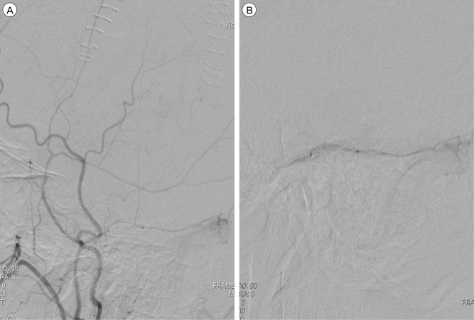
Fig. 2 (A) External carotid artery angiography identifies the dural arteriovenous fistula vascularized by the petrosqumosal branch of left middle meningeal artery (MMA) and drained into the transverse-sigmoid sinus. (B) Intraoperative angiogram of MMA indicates the microcatheter superselectively advanced into the petrosqumosal branch to eliminate the feeder and arteriovenous shunting.
Reference
-
1. Bansal H, Chaudhary A, Mahajan A, Paul B. Acute subdural hematoma secondary to cerebral venous sinus thrombosis: Case report and review of literature. Asian J Neurosurg. 2016; Apr-Jun. 11(2):177–180. PMID: 27057237.
Article2. Choi HJ, Cho CW. Anterior cranial fossa dural arteriovenous fistula presenting as subdural hematoma. J Korean Neurosurg Soc. 2010; 2. 47(2):155–157. PMID: 20224719.3. Daniels DJ, Vellimana AK, Zipfel GJ, Lanzino G. Intracranial hemorrhage from dural arteriovenous fistulas: clinical features and outcome. Neurosurg Focus. 2013; 5. 34(5):E15. PMID: 23634919.
Article4. Gandhi D, Chen J, Pearl M, Huang J, Gemmete JJ, Kathuria S. Intracranial dural arteriovenous fistulas: classification, imaging findings, and treatment. AJNR Am J Neuroradiol. 2012; 6. 33(6):1007–1013. PMID: 22241393.
Article5. Halbach VV, Higashida RT, Hieshima GB, Rosenblum M, Cahan L. Treatment of dural arteriovenous malformations involving the superior sagittal sinus. AJNR Am J Neuroradiol. 1988; Mar-Apr. 9(2):337–343. PMID: 3128082.6. Ito J, Imamura H, Kobayashi K, Tsuchida T, Sato S. Dural arteriovenous malformations of the base of the anterior cranial fossa. Neuroradiology. 1983; 3. 24(3):149–154. PMID: 6338411.
Article7. Kohyama S, Ishihara S, Yamane F, Kanazawa R, Ishihara H. Dural arteriovenous fistula presenting as an acute subdural hemorrhage that subsequently progressed to a chronic subdural hemorrhage: case report. Minim Invasive Neurosurg. 2009; 2. 52(1):36–38. PMID: 19247903.
Article8. Kominato Y, Matsui K, Hata Y, Matsui K, Kuwayama N, Ishizawa S, et al. Acute subdural hematoma due to arteriovenous malformation primarily in dura mater: a case report. Leg Med (Tokyo). 2004; 10. 6(4):256–260. PMID: 15363452.
Article9. Krishnaney AA, Rasmussen PA, Masaryk T. Bilateral tentorial subdural hematoma without subarachnoid hemorrhage secondary to anterior communicating artery aneurysm rupture: a case report and review of the literature. AJNR Am J Neuroradiol. 2004; Jun-Jul. 25(6):1006–1007. PMID: 15205138.10. Marquardt G, Weidauer S, Lanfermann H, Seifert V. Cerebral venous sinus thrombosis manifesting as bilateral subdural effusion: case report. Acta Neurol Scand. 2004; 109(6):425–428. PMID: 15147467.11. Missori P, Domenicucci M, Sassun TE, Tarantino R, Peschillo S. Alterations in the intracranial venous sinuses in spontaneous nontraumatic chronic subdural hematomas. J Clin Neurosci. 2013; 3. 20(3):389–393. PMID: 23219821.
Article12. Nomura S, Anegawa S, Nakagawa S, Tomokiyo M, Koga H, Hayashi T. Subarachnoid hemorrhage caused by dural arteriovenous fistula of the sphenobasal sinus: case report. Neurol Med Chir (Tokyo). 2002; 6. 42(6):255–258. PMID: 12116531.
Article13. Ogawa K, Oishi M, Mizutani T, Maejima S, Mori T. Dural arteriovenous fistula on the convexity presenting with pure acute subdural hematoma. Acta Neurol Belg. 2010; 6. 110(2):190–192. PMID: 20873450.14. Oh JS, Yoon SK, Oh HJ, Shim JJ, Bae HG, Lee KS. Endovascular treatment of dural arteriovenous fistulas: single center experience. J Korean Neurosurg Soc. 2016; 1. 59(1):17–25. PMID: 26885282.
Article15. Pappas CTE, Zabramski JM, Sheiter AG. Iatrogenic arteriovenous fistula presenting as a recurrent subdural hematoma: case report. J Neurosurg. 1992; 1. 76(1):134–136. PMID: 1727151.16. Peng T, Liu A, Jia J, Jiang C, Li Y, Wu Z, et al. Risk factors for dural arteriovenous fistula intracranial hemorrhage. J Clin Neurosci. 2014; 5. 21(5):769–772. PMID: 24291477.
Article17. Signorelli F, Della Pepa GM, Sabatino G, Marchese E, Maira G, Puca A, et al. Diagnosis and management of dural arteriovenous fistulas: a 10 years single-center experience. Clin Neurol Neurosurg. 2015; 1. 128(1):123–129. PMID: 25496935.
Article18. Suzuki K, Kamezaki T, Tsuboi K, Kobayashi E. Dural cavernous angioma causing acute subdural hemorrhage: case report. Neurol Med Chir (Tokyo). 1996; 8. 36(8):580–582. PMID: 8831201.
Article19. Takahashi S, Shinoda J, Hayashi T. Cerebral venous sinus thrombosis in an adult patient presenting as headache and acute subdural hematoma. J Stroke Cerebrovasc Dis. 2012; 5. 21(4):338–340. PMID: 21185743.
Article20. Van Rooij WJ, Sluzewski M, Beute GN. Dural arteriovenous fistulas with cortical venous drainage: incidence, clinical presentation, and treatment. AJNR Am J Neuroradiol. 2007; 4. 28(4):651–655. PMID: 17416815.
- Full Text Links
-
- Actions
-
Cited
- CITED
-
- Close
- Share
-
- Similar articles
-
- Spontaneous Intracranial Hypotension, a Possible Cause of Chronic Subdural Hematoma
- Intracranial Dural Arteriovenous Malfirmation: Report of Three Cases
- Spontaneous Spinal Subdural and Subarachnoid Hemorrhage with Concomitant Intracerebral Hemorrhage: A Case Report
- Intraoperative Development of Contralateral Subdural Hematoma during Evacuation of Acute Subdural Hematoma: Case Report
- Spinal Epidural Arteriovenous Fistula Presented with Subdural Hematoma: a Case of Transarterial Embolization Using NBCA
