J Korean Med Sci.
2017 Mar;32(3):552-555. 10.3346/jkms.2017.32.3.552.
Unsuspected Duplicated Gallbladder in a Patient Presenting with Acute Cholecystitis
- Affiliations
-
- 1Department of Surgery, Gyeongsang National University Hospital, Gyeongsang National University Postgraduate School of Medicine, Jinju, Korea. drjcy@hanmail.net
- 2Department of Pathology, Gyeongsang National University Hospital, Gyeongsang National University Postgraduate School of Medicine, Jinju, Korea.
- KMID: 2366655
- DOI: http://doi.org/10.3346/jkms.2017.32.3.552
Abstract
- Duplicated gallbladder (GB) is a rare congenital disease. Surgical management of a duplicated GB needs special care because of concurrent bile duct anomalies and the risk of injuring adjacent arteries during surgery. An 80-year-old man visited an emergency room with right upper quadrant abdominal pain. Computed tomography (CT) revealed cholecystitis with a 2-bodied GB. Because of this unusual finding, magnetic resonance choledochopancreatography was performed to detect possible biliary anomalies. The 2 GB bodies were unified at the neck with a common cystic duct, a so-called V-shaped duplicated GB. The patient's right posterior hepatic duct joined the common bile duct (CBD) near the cystic duct. The patient underwent laparoscopic cholecystectomy without adjacent organ injury, and was discharged uneventfully. Surgeons should carefully evaluate the patient preoperatively and select adequate surgical procedures in patients with suspected duplicated GB because of the risk of concurrent biliary anomalies.
MeSH Terms
Figure
-
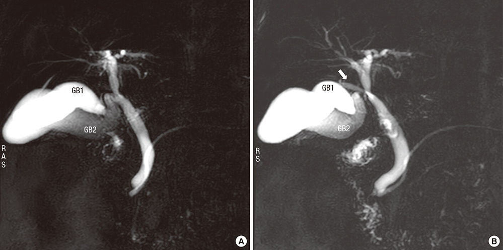
Fig. 1 Magnetic resonance choledochopancreatography reveals V-shpaed duplicated GB with right hepatic duct anomaly. (A) GB 1 was inserted into the neck of the other GB (GB 2). (B) The right posterior bile duct (arrow) joined the CBD near the common cystic duct. GB = gallbladder, CBD = common bile duct.
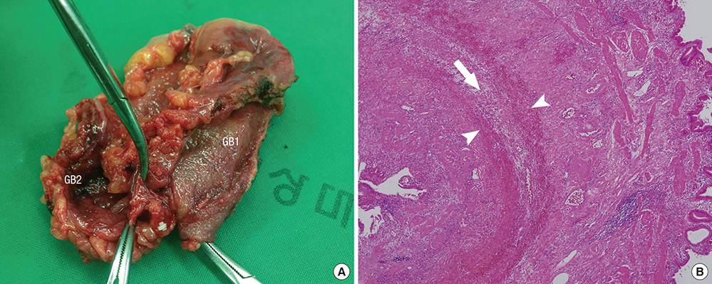
Fig. 2 Macroscopic view (A) of the specimen and microscopic features of the duplicated GB (B). The GB body shows separation of the lamina propria and muscle layer (arrowhead). Perimuscular connective tissue (arrow) is shared between the GB 2 bodies with H & E staining (× 40). GB = gallbladder, H & E = hematoxylin and eosin.

Fig. 3 Classification of duplicated GB. Adapted from Harlaftis et al. (1). GB = gallbladder.
Reference
-
1. Harlaftis N, Gray SW, Skandalakis JE. Multiple gallbladders. Surg Gynecol Obstet. 1977; 145:928–934.2. Shiba H, Misawa T, Ito R, Ohki K, Igarashi T, Yanaga K. Duplicated gallbladder. Int Surg. 2014; 99:77–78.3. Paraskevas GK, Raikos A, Ioannidis O, Papaziogas B. Duplicated gallbladder: surgical application and review of the literature. Ital J Anat Embryol. 2011; 116:61–66.4. Singh B, Ramsaroop L, Allopi L, Moodley J, Satyapal KS. Duplicate gallbladder: an unusual case report. Surg Radiol Anat. 2006; 28:654–657.5. Gocmen R, Yesilkaya Y. Imaging findings of gallbladder duplication due to two cases: case report and review of literature. Med Ultrason. 2012; 14:358–360.6. Botsford A, McKay K, Hartery A, Hapgood C. MRCP imaging of duplicate gallbladder: a case report and review of the literature. Surg Radiol Anat. 2015; 37:425–429.7. Huang BK, Chess MA. Cholecystitis of a duplicated gallbladder complicated by a cholecystoenteric fistula. Pediatr Radiol. 2009; 39:385–388.8. Ozaki N, Hashimoto D, Ikuta Y, Chikamoto A, Takamori H, Baba H. Definitive diagnosis of a duplicate gallbladder can only be made intraoperatively: report of a case. Clin J Gastroenterol. 2014; 7:338–341.9. Han JY, Jeong S, Lee DH. Percutaneous papillary large balloon dilation during percutaneous cholangioscopic lithotripsy for the treatment of large bile-duct stones: a feasibility study. J Korean Med Sci. 2015; 30:278–282.10. Reinisch A, Brandt L, Fuchs KH. Gallbladder duplication--laparoscopic cholecystectomy 17 years after open cholecystectomy. Zentralbl Chir. 2009; 134:576–579.11. Causey MW, Miller S, Fernelius CA, Burgess JR, Brown TA, Newton C. Gallbladder duplication: evaluation, treatment, and classification. J Pediatr Surg. 2010; 45:443–446.12. Walbolt TD, Lalezarzadeh F. Laparoscopic management of a duplicated gallbladder: a case study and anatomic history. Surg Laparosc Endosc Percutan Tech. 2011; 21:e156–8.13. Shirahane K, Yamaguchi K, Ogawa T, Shimizu S, Yokohata K, Mizumoto K, Tanaka M. Gallbladder duplication successfully removed laparoscopically using endoscopic nasobiliary tube. Surg Endosc. 2003; 17:1156.
- Full Text Links
-
- Actions
-
Cited
- CITED
-
- Close
- Share
-
- Similar articles
-
- Beneficial Effect of Cholecystography following PGBD for Complicated Acute Cholecystitis: Detection of Unsuspected CBD Stone
- Endoscopic Transpapillary Gallbladder Stenting for Acute Cholecystitis in a Patient with Metastatic Pancreatic Cancer
- Acute Cholecystitis Associated with Gallbladder Metastasis of Gastric Cancer
- Contracted Chronic Cholecystitis with Gallstone
- Necrotizing Gallbladder Torsion Masking as Acalculous Cholecystitis: A Review of Two Cases Treated with Successful Laparoscopic Cholecystectomy
