Estimation of Intraocular Lens Power Calculation after Myopic Corneal Refractive Surgery: Using Corneal Height in Anterior Segment Optical Coherence Tomography
- Affiliations
-
- 1Department of Ophthalmology, Seoul National University College of Medicine, Seoul, Korea. kmk9@snu.ac.kr
- 2Laboratory of Ocular Regenerative Medicine and Immunology, Seoul Artificial Eye Center, Seoul National University Hospital Clinical Research Institute, Seoul, Korea.
- KMID: 2363759
- DOI: http://doi.org/10.3341/kjo.2015.29.3.195
Abstract
- PURPOSE
To investigate the feasibility of estimating effective lens position (ELP) and calculating intraocular lens power using corneal height (CH), as measured using anterior segment optical coherence tomography (AS-OCT), in patients who have undergone corneal refractive surgery.
METHODS
This study included 23 patients (30 eyes) who have undergone myopic corneal refractive surgery and subsequent successful cataract surgery. The CH was measured with AS-OCT, and the measured ELP (ELP(m)) was calculated. Intraocular lens power, which could achieve actual emmetropia (P(real)), was determined with medical records. Estimated ELP (ELP(est)) was back-calculated using P(real), axial length, and keratometric value through the SRK/T formula. After searching the best-fit regression formula between ELP(m) and ELP(est), converted ELP and intraocular lens power (ELP(conv), P(conv)) were obtained and then compared to ELP(est) and P(real), respectively. The proportion of eyes within a defined error was investigated.
RESULTS
Mean CH, ELP(est), and ELP(m) were 3.71 +/- 0.23, 7.74 +/- 1.09, 5.78 +/- 0.26 mm, respectively. The ELP(m) and ELP(est) were linearly correlated (ELP(est) = 1.841 x ELP(m) - 2.018, p = 0.023, R = 0.410) and ELP(conv) and P(conv) agreed well with ELP(est) and P(real), respectively. Eyes within +/-0.5, +/-1.0, +/-1.5, and +/-2.0 diopters of the calculated P(conv), were 23.3%, 66.6%, 83.3%, and 100.0%, respectively.
CONCLUSIONS
Intraocular lens power calculation using CH measured with AS-OCT shows comparable accuracy to several conventional methods in eyes following corneal refractive surgery.
Keyword
MeSH Terms
Figure
-

Fig. 1 An anterior segment optical coherence tomography image. The measured corneal height (CHm) was defined as the distance between the posterior corneal surface and the intersection between the perpendicular lines.

Fig. 2 Linear regression analysis of measured effective lens position (ELPm) and the estimated effective lens position (ELPest).
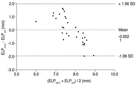
Fig. 3 Bland-Altman plot showing differences between measured effective lens position (ELPm) and estimated effective lens position (ELPest). Lines showing the mean difference and the ±1.96 standard deviation (SD) limits are also shown.
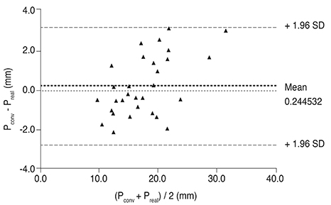
Fig. 4 Bland-Altman plot showing differences between the converted intraocular lens power (Pconv) and the actual intraocular lens power (Preal). Lines showing the mean difference and the ±1.96 standard deviation (SD) limits are also shown.
Reference
-
1. Holladay JT. Consultations in refractive surgery. Refract Corneal Surg. 1989; 5:202–203.2. Hoffer KJ. Intraocular lens power calculation for eyes after refractive keratotomy. J Refract Surg. 1995; 11:490–493.3. Seitz B, Langenbucher A. Intraocular lens power calculation in eyes after corneal refractive surgery. J Refract Surg. 2000; 16:349–361.4. Feiz V, Mannis MJ, Garcia-Ferrer F, et al. Intraocular lens power calculation after laser in situ keratomileusis for myopia and hyperopia: a standardized approach. Cornea. 2001; 20:792–797.5. Speicher L. Intra-ocular lens calculation status after corneal refractive surgery. Curr Opin Ophthalmol. 2001; 12:17–29.6. Rosa N, Capasso L, Romano A. A new method of calculating intraocular lens power after photorefractive keratectomy. J Refract Surg. 2002; 18:720–724.7. Haigis W. Corneal power after refractive surgery for myopia: contact lens method. J Cataract Refract Surg. 2003; 29:1397–1411.8. Ferrara G, Cennamo G, Marotta G, Loffredo E. New formula to calculate corneal power after refractive surgery. J Refract Surg. 2004; 20:465–471.9. Wang L, Booth MA, Koch DD. Comparison of intraocular lens power calculation methods in eyes that have undergone LASIK. Ophthalmology. 2004; 111:1825–1831.10. Latkany RA, Chokshi AR, Speaker MG, et al. Intraocular lens calculations after refractive surgery. J Cataract Refract Surg. 2005; 31:562–570.11. Camellin M, Calossi A. A new formula for intraocular lens power calculation after refractive corneal surgery. J Refract Surg. 2006; 22:187–199.12. Jarade EF, Abi Nader FC, Tabbara KF. Intraocular lens power calculation following LASIK: determination of the new effective index of refraction. J Refract Surg. 2006; 22:75–80.13. Masket S, Masket SE. Simple regression formula for intraocular lens power adjustment in eyes requiring cataract surgery after excimer laser photoablation. J Cataract Refract Surg. 2006; 32:430–434.14. Savini G, Barboni P, Zanini M. Intraocular lens power calculation after myopic refractive surgery: theoretical comparison of different methods. Ophthalmology. 2006; 113:1271–1282.15. Olsen T. Calculation of intraocular lens power: a review. Acta Ophthalmol Scand. 2007; 85:472–485.16. Haigis W. Intraocular lens calculation after refractive surgery for myopia: Haigis-L formula. J Cataract Refract Surg. 2008; 34:1658–1663.17. Saiki M, Negishi K, Kato N, et al. A new central-peripheral corneal curvature method for intraocular lens power calculation after excimer laser refractive surgery. Acta Ophthalmol. 2013; 91:e133–e139.18. Holladay JT, Prager TC, Chandler TY, et al. A three-part system for refining intraocular lens power calculations. J Cataract Refract Surg. 1988; 14:17–24.19. Retzlaff JA, Sanders DR, Kraff MC. Development of the SRK/T intraocular lens implant power calculation formula. J Cataract Refract Surg. 1990; 16:333–340.20. Hoffer KJ. The Hoffer Q formula: a comparison of theoretic and regression formulas. J Cataract Refract Surg. 1993; 19:700–712.21. Ho JD, Liou SW, Tsai RJ, Tsai CY. Estimation of the effective lens position using a rotating Scheimpflug camera. J Cataract Refract Surg. 2008; 34:2119–2127.22. Aristodemou P, Knox Cartwright NE, Sparrow JM, Johnston RL. Formula choice: Hoffer Q, Holladay 1, or SRK/T and refractive outcomes in 8108 eyes after cataract surgery with biometry by partial coherence interferometry. J Cataract Refract Surg. 2011; 37:63–71.23. Doors M, Berendschot TT, de Brabander J, et al. Value of optical coherence tomography for anterior segment surgery. J Cataract Refract Surg. 2010; 36:1213–1229.24. Yang R, Yeh A, George MR, et al. Comparison of intraocular lens power calculation methods after myopic laser refractive surgery without previous refractive surgery data. J Cataract Refract Surg. 2013; 39:1327–1335.25. Arce CG, Soriano ES, Weisenthal RW, et al. Calculation of intraocular lens power using Orbscan II quantitative area topography after corneal refractive surgery. J Refract Surg. 2009; 25:1061–1074.26. Olsen T, Corydon L, Gimbel H. Intraocular lens power calculation with an improved anterior chamber depth prediction algorithm. J Cataract Refract Surg. 1995; 21:313–319.27. Tomlinson A, Leighton DA. Ocular dimensions in the heredity of angle-closure glaucoma. Br J Ophthalmol. 1973; 57:475–486.28. Olsen T. Prediction of the effective postoperative (intraocular lens) anterior chamber depth. J Cataract Refract Surg. 2006; 32:419–424.29. Nishimura R, Negishi K, Dogru M, et al. Effect of age on changes in anterior chamber depth and volume after laser in situ keratomileusis. J Cataract Refract Surg. 2009; 35:1868–1872.30. Doors M, Cruysberg LP, Berendschot TT, et al. Comparison of central corneal thickness and anterior chamber depth measurements using three imaging technologies in normal eyes and after phakic intraocular lens implantation. Graefes Arch Clin Exp Ophthalmol. 2009; 247:1139–1146.31. Yi JH, Hong S, Seong GJ, et al. Anterior chamber measurements by pentacam and AS-OCT in eyes with normal open angles. Korean J Ophthalmol. 2008; 22:242–245.32. McCarthy M, Gavanski GM, Paton KE, Holland SP. Intraocular lens power calculations after myopic laser refractive surgery: a comparison of methods in 173 eyes. Ophthalmology. 2011; 118:940–944.33. Sonego-Krone S, Lopez-Moreno G, Beaujon-Balbi OV, et al. A direct method to measure the power of the central cornea after myopic laser in situ keratomileusis. Arch Ophthalmol. 2004; 122:159–166.34. Tang M, Wang L, Koch DD, et al. Intraocular lens power calculation after previous myopic laser vision correction based on corneal power measured by Fourier-domain optical coherence tomography. J Cataract Refract Surg. 2012; 38:589–594.
- Full Text Links
-
- Actions
-
Cited
- CITED
-
- Close
- Share
-
- Similar articles
-
- Corneal Power Estimation Using Orbscan II Videokeratography in Eyes With Previous Corneal Refractive Surgeries
- Anterior Segment Swept-Source Optical Coherence Tomography–based Assessment of Corneal Refractive Profiles in Stevens-Johnson Syndrome/Toxic Epidermal Necrolysis Patients: A Controlled Comparative Study
- Keratometry and Computerized Videokeratography in Determining Intraocular Lens Calculation
- Inter-eye Difference of the Intraocular Lens Power Calculation in a Case with Bilateral Posterior Keratoconus
- The Effect of Refractive Power on Retinal Volume Measurement Using Spectral Domain Optical Coherence Tomography
