First Report on Familial Hemophagocytic Lymphohistiocytosis with an Abnormal Immunophenotype and T Cell Monoclonality in Korea
- Affiliations
-
- 1Department of Laboratory Medicine & Genetics, Samsung Medical Center, Sungkyunkwan University School of Medicine, Seoul, Korea. sunnyhk@skku.edu, heejinkim@skku.edu
- 2Department of Pediatrics, Samsung Medical Center, Sungkyunkwan University School of Medicine, Seoul, Korea.
- 3Department of Pathology, Samsung Medical Center, Sungkyunkwan University School of Medicine, Seoul, Korea.
- KMID: 2363167
- DOI: http://doi.org/10.3343/alm.2015.35.1.155
Abstract
- No abstract available.
MeSH Terms
-
Bone Marrow/metabolism/pathology
DNA Mutational Analysis
Gene Rearrangement, T-Lymphocyte
Humans
Immunophenotyping
Infant
Lymphohistiocytosis, Hemophagocytic/*diagnosis
Male
Membrane Proteins/chemistry/genetics
Polymorphism, Single-Stranded Conformational
Republic of Korea
T-Lymphocytes/immunology/*metabolism
Membrane Proteins
Figure
-
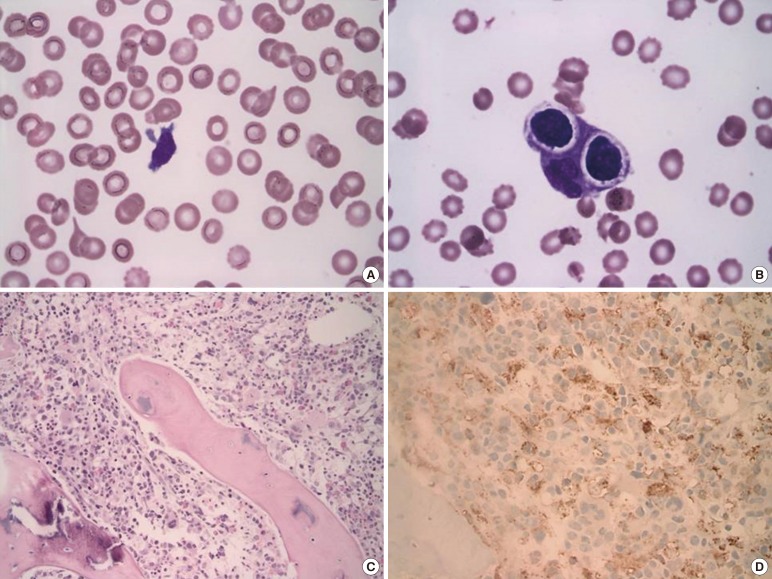
Fig. 1 Peripheral blood smear, bone marrow biopsy and aspirate. (A) Pancytopenia and atypical lymphoid cells were observed in peripheral blood smear (Wright-Giemsa stain; magnification, ×1,000). (B) Hemophagocytic histiocytes were frequently observed in bone marrow aspiration (Wright-Giemsa stain; magnification, ×1,000). (C) Histiocytes were high in number in the biopsy section (Hematoxylin-Eosin stain; magnification ×200). (D) CD68 immunization (magnification, ×400).
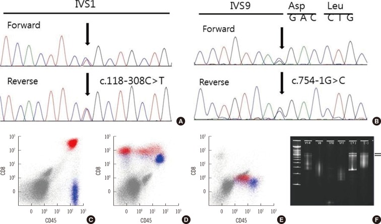
Fig. 2 Sequencing analysis, flow cytometry results, and single-strand conformational polymorphism (SSCP) analysis of the T cell receptor by using paraffin-embedded tissue samples. (A) Compound heterozygous mutations in the UNC13D gene were observed: C to T substitution at nucleotide -308 of intron 1, relative to the cDNA positioned between nucleotides 117 and 118 (118-308C>T). (B) G to C substitution at nucleotide -1 of intron 9, relative to the cDNA positioned between nucleotides 753 and 754 (754-1G>C). The CD3+/CD8+ T cells (red). (C) showed variable downregulation of CD5 or CD7 (lack of expression) compared with CD3+/CD4+ cells (blue, presumably normal T cells; D and E). (F) DNA was extracted from paraffin-embedded bone marrow tissue. Consensus primer for Vγ1-8, Vγ9-11 was used for amplification. PCR products were analyzed by SSCP, which separated DNA fragments according to nucleotide sequence. Well-defined, distinct bands were considered evidence of monoclonality. The two black arrows indicate the distinct monoclonal band at the level of the lowest smear in the polyclonal control (-). Monoclonality was observed in Vγ1-8, and polyclonality was observed in Vγ9-11.
Reference
-
1. McCall CM, Mudali S, Arceci RJ, Small D, Fuller S, Gocke CD, et al. Flow cytometric findings in hemophagocytic lymphohistiocytosis. Am J Clin Pathol. 2012; 137:786–794. PMID: 22523218.
Article2. Ahn JS, Rew SY, Shin MG, Kim HR, Yang DH, Cho D, et al. Clinical significance of clonality and Epstein-Barr virus infection in adult patients with hemophagocytic lymphohistiocytosis. Am J Hematol. 2010; 85:719–722. PMID: 20652965.
Article3. Karandikar NJ, Kroft SH, Yegappan S, Rogers BB, Aquino VM, Lee KM, et al. Unusual immunophenotype of CD8+ T cells in familial hemophagocytic lymphohistiocytosis. Blood. 2004; 104:2007–2009. PMID: 15205266.
Article4. Seo JY, Song JS, Lee KO, Won HH, Kim JW, Kim SH, et al. Founder effects in two predominant intronic mutations of UNC13D, c.118-308C>T and c.754-1G>C underlie the unusual predominance of type 3 familial hemophagocytic lymphohistiocytosis (FHL3) in Korea. Ann Hematol. 2013; 92:357–364. PMID: 23180437.5. Signoretti S, Murphy M, Cangi MG, Puddu P, Kadin ME, Loda M. Detection of clonal T-cell receptor gamma gene rearrangements in paraffin-embedded tissue by polymerase chain reaction and nonradioactive single-strand conformational polymorphism analysis. Am J Pathol. 1999; 154:67–75. PMID: 9916920.6. Toga A, Wada T, Sakakibara Y, Mase S, Araki R, Tone Y, et al. Clinical significance of cloned expansion and CD5 down-regulation in Epstein-Barr Virus (EBV)-infected CD8+ T lymphocytes in EBV-associated hemophagocytic lymphohistiocytosis. J Infect Dis. 2010; 201:1923–1932. PMID: 20443735.7. Lin MT, Chang HM, Huang CJ, Chen WL, Lin CY, Chuang SS. Massive expansion of EBV+ monoclonal T cells with CD5 down regulation in EBV-associated haemophagocytic lymphohistiocytosis. J Clin Pathol. 2007; 60:101–103. PMID: 17213357.
Article8. Wada T, Sakakibara Y, Nishimura R, Toma T, Ueno Y, Horita S, et al. Down-regulation of CD5 expression on activated CD8+ T cells in familial hemophagocytic lymphohistiocytosis with perforin gene mutations. Hum Immunol. 2013; 74:1579–1585. PMID: 24051121.
Article9. Imashuku S, Hibi S, Tabata Y, Itoh E, Hashida T, Tsunamoto K, et al. Outcome of clonal hemophagocytic lymphohistiocytosis: analysis of 32 cases. Leuk Lymphoma. 2000; 37:577–584. PMID: 11042518.
Article10. Ishii E, Kimura N, Kato K, Sako M, Nagano M, Nakagawa A, et al. Clonal change of infiltrating T-cells in children with familial hemophagocytic lymphohistiocytosis: possible association with Epstein-Barr virus infection. Cancer. 1999; 85:1636–1643. PMID: 10193957.
- Full Text Links
-
- Actions
-
Cited
- CITED
-
- Close
- Share
-
- Similar articles
-
- Flow Cytometric Analysis of T Cells in Hemophagocytic Lymphohistiocytosis
- A Case of Secondary Precursor B-cell Acute Lymphoblastic Leukemia Occurring after Treatment of Hemophagocytic Lymphohistiocytosis
- EBV-elicited familial hemophagocytic lymphohistiocytosis
- Hemophagocytic Lymphohistiocytosis in Adults: Overview, Diagnosis, and Treatment
- Two Cases of Hemophagocytic Lymphohistiocytosis Following Kikuchi's Disease
