Detectability and Usefulness of Automated Whole Breast Ultrasound in Patients with Suspicious Microcalcifications on Mammography: Comparison with Handheld Breast Ultrasound
- Affiliations
-
- 1Department of Radiology, Dongtan Sacred Heart Hospital, Hallym University Medical Center, Hwaseong, Korea.
- 2Department of Radiology, Seoul St. Mary's Hospital, The Catholic University of Korea College of Medicine, Seoul, Korea. rad-ksh@catholic.ac.kr
- 3Department of General Surgery, Seoul St. Mary's Hospital, The Catholic University of Korea College of Medicine, Seoul, Korea.
- KMID: 2362930
- DOI: http://doi.org/10.4048/jbc.2016.19.4.429
Abstract
- PURPOSE
The purpose of this study was to prospectively evaluate the detectability and usefulness of automated whole breast ultrasound (AWUS) and to compare it with handheld breast ultrasound (HHUS) in cases with suspicious microcalcifications identified by mammography.
METHODS
Forty-two patients with 43 suspicious microcalcifications (25 malignant and 18 benign) detected by mammography underwent AWUS, HHUS, and histol-ogic examination. With knowledge of the mammographic findings, HHUS was performed to assess the visibility of the microcalcifications and the presence of associated masses or ductal changes. Two radiologists reviewed the AWUS images in consensus using the same methods employed for HHUS. Detectability of AWUS was compared with that of HHUS and was correlated with histologic and mammographic findings.
RESULTS
Of the 43 lesions, 32 (74.4%) were detectable by AWUS and 31 (72.1%) by HHUS. No significant differences in sensitivity were found between the two methods (p=0.998). AWUS detected 96% (24/25) of malignant microcalcifications and 44.4% (8/18) of benign microcalcifications. AWUS was more successful in the detection of malignant vs. benign lesions (96.0% vs. 44.4%, p=0.002), lesions >10 mm vs. ≤10 mm in size (86.7% [26/30] vs. 46.2% [6/13], p=0.009), lesions with a fine pleomorphic or linear shape vs. a round or amorphous or coarse heterogeneous shape (94.7% [18/19] vs. 58.3% [14/24], p=0.021), and lesions associated with a mass or architectural distortion vs. without obvious changes on mammography (100% [19/19] vs. 54.2% [13/24], p=0.022).
CONCLUSION
Detectability of AWUS was comparable to that of HHUS in cases where suspicious microcalcifications were identified on mammography. Therefore, AWUS might be helpful in the performance of ultrasound-guided percutaneous procedures for highly suspicious microcalcifications.
MeSH Terms
Figure
-
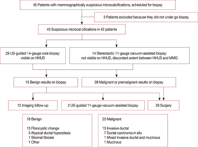
Figure 1 Flow chart shows the study population, inclusion and exclusion criteria, and pathologic findings. Three patients with benign results on biopsy underwent 11-gauge vacuum-assisted biopsy or surgical excision due to patient anxiety. US=ultrasound; HHUS=handheld breast ultrasound; MMG=mammography.
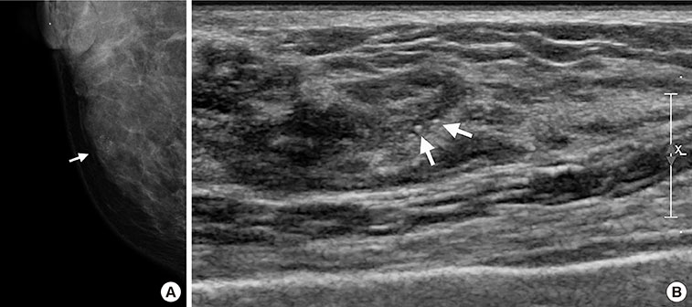
Figure 2 A 42-year-old woman with fibrocystic disease in the right breast. (A) Spot magnification mammogram shows grouped punctate microcalcifications in inner breast (arrow). (B) Handheld ultrasound (US) shows hyperechoic microcalcifications (arrows). These microcalcifications were not seen by automated whole breast US. This may be due to the lower resolution of automated whole breast US.
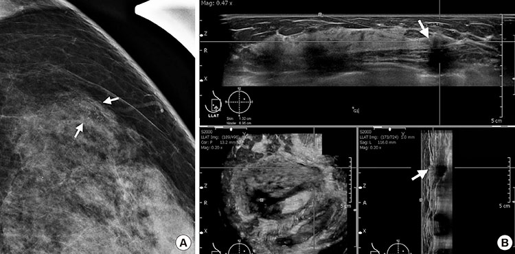
Figure 3 A 49-year-old woman with fibrocystic disease in the left breast. (A) Spot magnification mammogram shows grouped punctate or amorphous microcalcifications in outer breast (arrows). (B) Automated whole breast ultrasound (US) shows hyperechoic microcalcifications within the hypoechoic area (arrows). These microcalcifications were not detected by handheld US. This may be due to the operator dependency of handheld breast US.
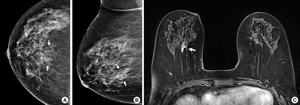
Figure 4 A 58-year-old woman with ductal carcinoma in situ in the right breast. (A, B) Craniocaudal and mediolateral oblique views of mammogram shows fine pleomorphic microcalcifications in deep central breast (arrows). (C) Axial T1-weighted contrast-enhanced magnetic resonance image shows an irregular enhancing mass in central posterior portion of right breast (arrow). These microcalcifications were not seen by hand held ultrasound (US) and automated whole breast US.
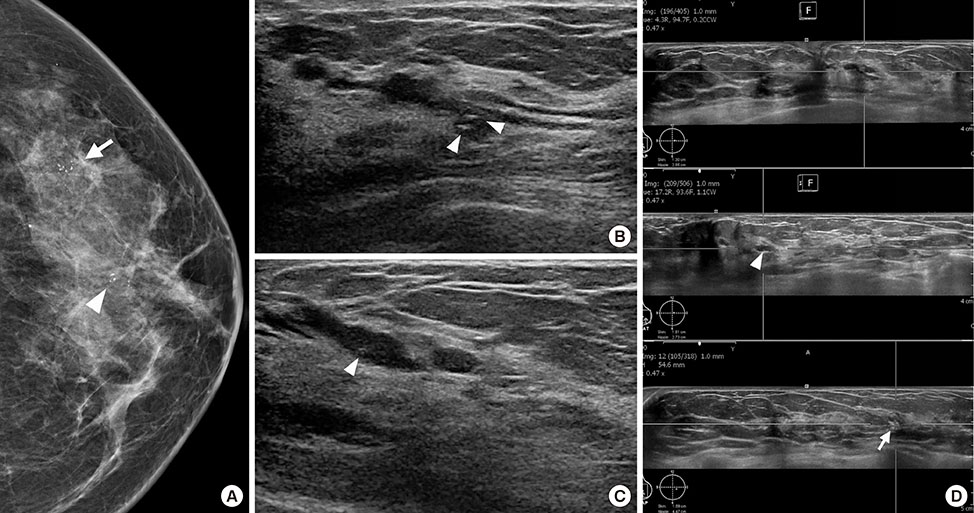
Figure 5 A 43-year-old woman with ductal carcinoma in situ in the left breast. (A) Craniocaudal view of mammogram shows segmental fine pleomorphic microcalcifications in central (arrowhead) and peripheral outer breast (arrow). (B, C) Handheld ultrasound (US) shows an intraductal hypoechoic mass with microcalcifications (arrowheads), which matched with central microcalcifications on mammogram. (D) Automated whole breast US shows segmental ductal dilatation with intraductal microcalcifications (arrowhead), which matched with central microcalcifications on mammogram. Grouped microcalcifications (arrow) in peripheral breast are found, which matched with peripheral microcalcifications on mammogram.
Reference
-
1. Sickles EA. Breast calcifications: mammographic evaluation. Radiology. 1986; 160:289–293.
Article2. Sickles EA. Mammographic features of 300 consecutive nonpalpable breast cancers. AJR Am J Roentgenol. 1986; 146:661–663.
Article3. Ciatto S, Cataliotti L, Distante V. Nonpalpable lesions detected with mammography: review of 512 consecutive cases. Radiology. 1987; 165:99–102.
Article4. Meyer JE, Kopans DB, Stomper PC, Lindfors KK. Occult breast abnormalities: percutaneous preoperative needle localization. Radiology. 1984; 150:335–337.
Article5. Meyer JE, Eberlein TJ, Stomper PC, Sonnenfeld MR. Biopsy of occult breast lesions: analysis of 1261 abnormalities. JAMA. 1990; 263:2341–2343.
Article6. Orel SG, Kay N, Reynolds C, Sullivan DC. BI-RADS categorization as a predictor of malignancy. Radiology. 1999; 211:845–850.
Article7. Moon WK, Im JG, Koh YH, Noh DY, Park IA. US of mammographically detected clustered microcalcifications. Radiology. 2000; 217:849–854.
Article8. Gufler H, Buitrago-Téllez CH, Madjar H, Allmann KH, Uhl M, Rohr-Reyes A. Ultrasound demonstration of mammographically detected microcalcifications. Acta Radiol. 2000; 41:217–221.
Article9. Hashimoto BE, Kramer DJ, Picozzi VJ. High detection rate of breast ductal carcinoma in situ calcifications on mammographically directed high-resolution sonography. J Ultrasound Med. 2001; 20:501–508.
Article10. Cheung YC, Wan YL, Chen SC, Lui KW, Ng SH, Yeow KM, et al. Sonographic evaluation of mammographically detected microcalcifications without a mass prior to stereotactic core needle biopsy. J Clin Ultrasound. 2002; 30:323–331.
Article11. Soo MS, Baker JA, Rosen EL, Vo TT. Sonographically guided biopsy of suspicious microcalcifications of the breast: a pilot study. AJR Am J Roentgenol. 2002; 178:1007–1015.
Article12. Soo MS, Baker JA, Rosen EL. Sonographic detection and sonographically guided biopsy of breast microcalcifications. AJR Am J Roentgenol. 2003; 180:941–948.
Article13. Shipley JA, Duck FA, Goddard DA, Hillman MR, Halliwell M, Jones MG, et al. Automated quantitative volumetric breast ultrasound data-acquisition system. Ultrasound Med Biol. 2005; 31:905–917.
Article14. Tozaki M, Fukuma E. Accuracy of determining preoperative cancer extent measured by automated breast ultrasonography. Jpn J Radiol. 2010; 28:771–773.
Article15. Kim SH, Kang BJ, Choi BG, Choi JJ, Lee JH, Song BJ, et al. Radiologists' performance for detecting lesions and the interobserver variability of automated whole breast ultrasound. Korean J Radiol. 2013; 14:154–163.
Article16. Shin HJ, Kim HH, Cha JH, Park JH, Lee KE, Kim JH. Automated ultrasound of the breast for diagnosis: interobserver agreement on lesion detection and characterization. AJR Am J Roentgenol. 2011; 197:747–754.
Article17. Lin X, Wang J, Han F, Fu J, Li A. Analysis of eighty-one cases with breast lesions using automated breast volume scanner and comparison with handheld ultrasound. Eur J Radiol. 2012; 81:873–878.
Article18. Wang HY, Jiang YX, Zhu QL, Zhang J, Dai Q, Liu H, et al. Differentiation of benign and malignant breast lesions: a comparison between automatically generated breast volume scans and handheld ultrasound examinations. Eur J Radiol. 2012; 81:3190–3200.
Article19. Kotsianos-Hermle D, Hiltawsky KM, Wirth S, Fischer T, Friese K, Reiser M. Analysis of 107 breast lesions with automated 3D ultrasound and comparison with mammography and manual ultrasound. Eur J Radiol. 2009; 71:109–115.
Article20. Kotsianos-Hermle D, Wirth S, Fischer T, Hiltawsky KM, Reiser M. First clinical use of a standardized three-dimensional ultrasound for breast imaging. Eur J Radiol. 2009; 71:102–108.
Article21. An YY, Kim SH, Kang BJ. The image quality and lesion characterization of breast using automated whole-breast ultrasound: a comparison with handheld ultrasound. Eur J Radiol. 2015; 84:1232–1235.
Article
- Full Text Links
-
- Actions
-
Cited
- CITED
-
- Close
- Share
-
- Similar articles
-
- Reliability of automated versus handheld breast ultrasound examinations of suspicious breast masses
- Clinical Applications of Automated Breast Ultrasound: Screening for Breast Cancer
- Automated Breast Ultrasound
- Calcifications on Breast Ultrasonography
- Usefulness of Ultrasound-Guided Automated Core Biopsy of Nonpalpable Breast Lesions
