Selenium Concentration in Korean Patients with Thyroid Disease: a Preliminary Report
- Affiliations
-
- 1Division of Endocrinology and Metabolism, Department of Medicine, Thyroid Center, Samsung Medical Center, Sungkyunkwan University School of Medicine, Seoul, Korea. jaeh.chung@samsung.com
- KMID: 2362351
- DOI: http://doi.org/10.11106/ijt.2016.9.2.152
Abstract
- BACKGROUND AND OBJECTIVES
Selenium is an important trace element for thyroid hormone metabolism, and its deficiency can cause hypothyroidism. Serum selenium concentration is the best biomarker to reflect selenium intake and reserve, although other markers can reflect. Therefore, we preliminarily assessed serum and urine selenium concentrations in patients with thyroid disease compared to those of a healthy population. We also investigated the correlation between serum and urine selenium concentration, thyroid hormone and urinary iodine concentration (UIC).
MATERIALS AND METHODS
A total of 97 patients (32 men, 65 women, 52.4±14.7 years) with benign thyroid nodules or thyroid dysfunction who visited the Samsung Medical Center between 2008 and 2013 were included. Data for 175 healthy subjects provided by Lee et al. were used as the control. Serum T3, free T4, and thyroid stimulating hormone (TSH) were measured using commercialized RIA or IRMA kits. Serum/urine selenium and UIC were measured by inductively coupled plasma-mass spectrometry (ICP-MS).
RESULTS
Median serum selenium concentration was 110 µg/L (95% CI, 73-156). Median urine selenium concentration was 66.3 µg/gCr (95% CI, 28.7-283.5). Compared to 175 healthy subjects (serum 84 µg/L [95% CI, 30-144], urine 34.5 µg/gCr [95% CI, 0.8-107.2]), serum and urine selenium concentrations of patients with thyroid disease were significantly higher than those of healthy subjects (p<0.001). Serum selenium concentration was significantly correlated with urine selenium concentration after log transformation (r=0.88, p=0.022), but was not significantly correlated with UIC, T3, free T4 and TSH.
CONCLUSION
Selenium concentrations of patients with thyroid disease were significantly higher than those of healthy subjects. Serum selenium concentration was significantly correlated with urine selenium concentration.
Keyword
MeSH Terms
Figure
-
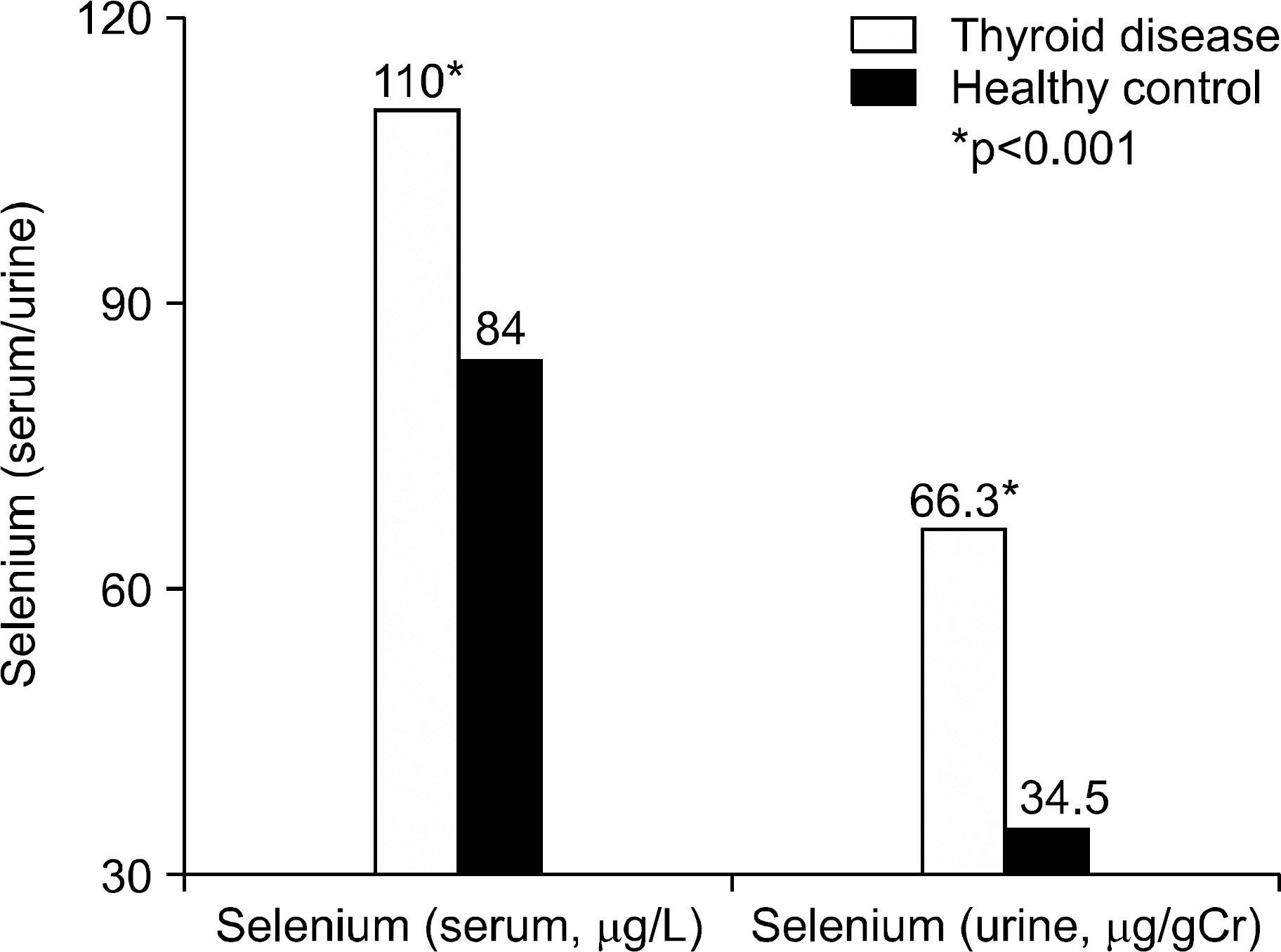
Fig. 1. Comparison of serum and urine selenium concen-trations of patients with thyroid disease to those of healthy subjects.
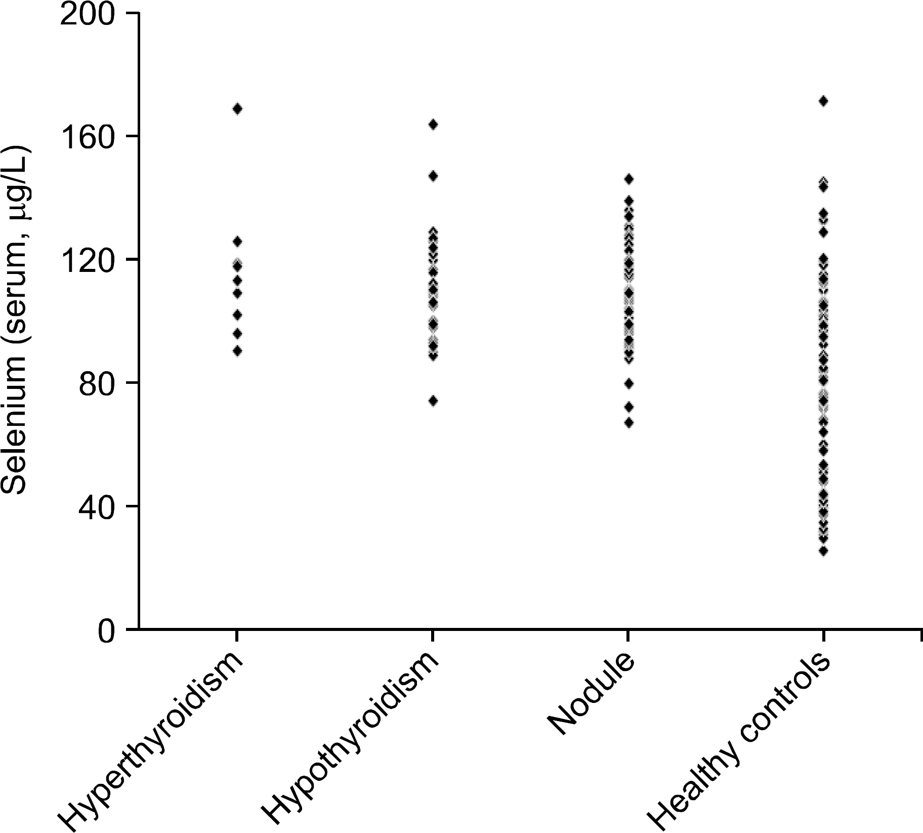
Fig. 2. Comparison of serum selenium concentrations of patients with various thyroid diseases to those of healthy subjects. Hyperthyroidism vs. controls; hypothyroidism vs. controls; nodule vs. controls, p<0.001.

Fig. 3. Comparison of urine selenium concentrations of patients with various thyroid diseases to those of healthy subjects. Hyperthyroidism vs. controls; hypothyroidism vs. controls; nodule vs. controls, p<0.001.

Fig. 4. Relationship between serum selenium and urine selenium after log transformation.
Cited by 1 articles
-
Treatment of Graves' Ophthalmopathy
Jeong Kyu Lee
Int J Thyroidol. 2019;12(2):91-96. doi: 10.11106/ijt.2019.12.2.91.
Reference
-
References
1. Duntas LH. Selenium and the thyroid: a close-knit connection. J Clin Endocrinol Metab. 2010; 95(12):5180–8.
Article2. Schomburg L. Selenium, selenoproteins and the thyroid gland: interactions in health and disease. Nat Rev Endocrinol. 2012; 8(3):160–71.
Article3. Rayman MP. Selenium and human health. Lancet. 2012; 379(9822):1256–68.
Article4. Effraimidis G, Wiersinga WM. Mechanisms in endocrinology: autoimmune thyroid disease: old and new players. Eur J Endocrinol. 2014; 170(6):R241–52.
Article5. Howie AF, Walker SW, Akesson B, Arthur JR, Beckett GJ. Thyroidal extracellular glutathione peroxidase: a potential regulator of thyroid-hormone synthesis. Biochem J. 1995; 308(Pt 3):713–7.
Article6. Schmutzler C, Mentrup B, Schomburg L, Hoang-Vu C, Herzog V, Kohrle J. Selenoproteins of the thyroid gland: expression, localization and possible function of glutathione peroxidase 3. Biol Chem. 2007; 388(10):1053–9.
Article7. Schomburg L, Kohrle J. On the importance of selenium and iodine metabolism for thyroid hormone biosynthesis and human health. Mol Nutr Food Res. 2008; 52(11):1235–46.
Article8. Salvatore D, Bartha T, Harney JW, Larsen PR. Molecular biological and biochemical characterization of the human type 2 selenodeiodinase. Endocrinology. 1996; 137(8):3308–15.
Article9. Rayman MP, Thompson AJ, Bekaert B, Catterick J, Galassini R, Hall E, et al. Randomized controlled trial of the effect of selenium supplementation on thyroid function in the elderly in the United Kingdom. Am J Clin Nutr. 2008; 87(2):370–8.
Article10. Olivieri O, Girelli D, Azzini M, Stanzial AM, Russo C, Ferroni M, et al. Low selenium status in the elderly influences thyroid hormones. Clin Sci (Lond). 1995; 89(6):637–42.
Article11. Duffield AJ, Thomson CD, Hill KE, Williams S. An estimation of selenium requirements for New Zealanders. Am J Clin Nutr. 1999; 70(5):896–903.
Article12. Thomson CD, McLachlan SK, Grant AM, Paterson E, Lillico AJ. The effect of selenium on thyroid status in a population with marginal selenium and iodine status. Br J Nutr. 2005; 94(6):962–8.
Article13. Hawkes WC, Keim NL, Diane Richter B, Gustafson MB, Gale B, Mackey BE, et al. High-selenium yeast supplementation in free-living North American men: no effect on thyroid hormone metabolism or body composition. J Trace Elem Med Biol. 2008; 22(2):131–42.
Article14. Gartner R, Gasnier BC, Dietrich JW, Krebs B, Angstwurm MW. Selenium supplementation in patients with autoimmune thyroiditis decreases thyroid peroxidase antibodies concentrations. J Clin Endocrinol Metab. 2002; 87(4):1687–91.
Article15. Duntas LH, Mantzou E, Koutras DA. Effects of a six month treatment with selenomethionine in patients with autoimmune thyroiditis. Eur J Endocrinol. 2003; 148(4):389–93.
Article16. Turker O, Kumanlioglu K, Karapolat I, Dogan I. Selenium treatment in autoimmune thyroiditis: 9-month follow-up with variable doses. J Endocrinol. 2006; 190(1):151–6.
Article17. Mazokopakis EE, Papadakis JA, Papadomanolaki MG, Batistakis AG, Giannakopoulos TG, Protopapadakis EE, et al. Effects of 12 months treatment with L-selenomethionine on serum anti-TPO levels in patients with Hashimoto's thyroiditis. Thyroid. 2007; 17(7):609–12.
Article18. Nacamulli D, Mian C, Petricca D, Lazzarotto F, Barollo S, Pozza D, et al. Influence of physiological dietary selenium supplementation on the natural course of autoimmune thyroiditis. Clin Endocrinol (Oxf). 2010; 73(4):535–9.
Article19. Negro R, Greco G, Mangieri T, Pezzarossa A, Dazzi D, Hassan H. The influence of selenium supplementation on postpartum thyroid status in pregnant women with thyroid peroxidase autoantibodies. J Clin Endocrinol Metab. 2007; 92(4):1263–8.
Article20. Bacic Vrca V, Skreb F, Cepelak I, Mayer L. Supplementation with antioxidants in the treatment of Graves' disease: the effect on the extracellular antioxidative parameters. Acta Pharm. 2004; 54(2):79–89.21. Marcocci C, Kahaly GJ, Krassas GE, Bartalena L, Prummel M, Stahl M, et al. Selenium and the course of mild Graves' orbitopathy. N Engl J Med. 2011; 364(20):1920–31.
Article22. Bleys J, Navas-Acien A, Guallar E. Serum selenium and diabetes in U.S. adults. Diabetes Care. 2007; 30(4):829–34.
Article23. Stranges S, Marshall JR, Natarajan R, Donahue RP, Trevisan M, Combs GF, et al. Effects of longterm selenium supplementation on the incidence of type 2 diabetes: a randomized trial. Ann Intern Med. 2007; 147(4):217–23.24. Stranges S, Laclaustra M, Ji C, Cappuccio FP, Navas-Acien A, Ordovas JM, et al. Higher selenium status is associated with adverse blood lipid profile in British adults. J Nutr. 2010; 140(1):81–7.
Article25. Combs GF Jr. Biomarkers of selenium status. Nutrients. 2015; 7(4):2209–36.
Article26. Sanz Alaejos M, Diaz Romero C. Urinary selenium concentrations. Clin Chem. 1993; 39(10):2040–52.
Article27. Lee SY, Oh HJ, Choi YH, Kim JW, Kim SH. Trace metal analysis using inductively coupled plasma-mass spectrometry (ICP-MS). Korean J Lab Med. 2004; 24(6):362–70.28. Lee JH, Ji OJ, Song MJ, Park HD, Kim HK, Kim SW, et al. Determination of urinary iodine concentration by inductively coupled plasma-mass spectrometry in thyroid cancer patients on low-iodine diet. Korean J Lab Med. 2010; 30(4):351–6.
Article29. Drutel A, Archambeaud F, Caron P. Selenium and the thyroid gland: more good news for clinicians. Clin Endocrinol (Oxf). 2013; 78(2):155–64.
Article30. Contempre B, Duale NL, Dumont JE, Ngo B, Diplock AT, Vanderpas J. Effect of selenium supplementation on thyroid hormone metabolism in an iodine and selenium deficient population. Clin Endocrinol (Oxf). 1992; 36(6):579–83.31. Contempre B, Dumont JE, Ngo B, Thilly CH, Diplock AT, Vanderpas J. Effect of selenium supplementation in hypothyroid subjects of an iodine and selenium deficient area: the possible danger of indiscriminate supplementation of iodine-deficient subjects with selenium. J Clin Endocrinol Metab. 1991; 73(1):213–5.32. Choi MK, Kang MH, Kim MH. The analysis of copper, selenium, and molybdenum contents in frequently consumed foods and an estimation of their daily intake in Korean adults. Biol Trace Elem Res. 2009; 128(2):104–17.
Article33. Combs GF Jr. Selenium in global food systems. Br J Nutr. 2001; 85(5):517–47.
Article34. Bleys J, Navas-Acien A, Guallar E. Selenium and diabetes: more bad news for supplements. Ann Intern Med. 2007; 147(4):271–2.
Article35. Combs GF Jr, Watts JC, Jackson MI, Johnson LK, Zeng H, Scheett AJ, et al. Determinants of selenium status in healthy adults. Nutr J. 2011; 10:75.
Article36. Hess SY. The impact of common micronutrient deficiencies on iodine and thyroid metabolism: the evidence from human studies. Best Pract Res Clin Endocrinol Metab. 2010; 24(1):117–32.
Article37. Olivieri O, Girelli D, Stanzial AM, Rossi L, Bassi A, Corrocher R. Selenium, zinc, and thyroid hormones in healthy subjects: low T3/T4 ratio in the elderly is related to impaired selenium status. Biol Trace Elem Res. 1996; 51(1):31–41.38. Thomson CD, Robinson MF, Campbell DR, Rea HM. Effect of prolonged supplementation with daily supplements of selenomethionine and sodium selenite on glutathione peroxidase activity in blood of New Zealand residents. Am J Clin Nutr. 1982; 36(1):24–31.
Article39. Thavarajah D, Vandenberg A, George GN, Pickering IJ. Chemical form of selenium in naturally selenium-rich lentils (Lens culinaris L.) from Saskatchewan. J Agric Food Chem. 2007; 55(18):7337–41.
Article40. Pedrosa LF, Motley AK, Stevenson TD, Hill KE, Burk RF. Fecal selenium excretion is regulated by dietary selenium intake. Biol Trace Elem Res. 2012; 149(3):377–81.
Article41. Hargreaves MK, Liu J, Buchowski MS, Patel KA, Larson CO, Schlundt DG, et al. Plasma selenium biomarkers in low income black and white americans from the southeastern United States. PLoS One. 2014; 9(1):): e84972.
Article42. Salvatore D, Tu H, Harney JW, Larsen PR. Type 2 iodothyronine deiodinase is highly expressed in human thyroid. J Clin Invest. 1996; 98(4):962–8.
Article43. Moreno-Reyes R, Suetens C, Mathieu F, Begaux F, Zhu D, Rivera MT, et al. Kashin-Beck osteoarthropathy in rural Tibet in relation to selenium and iodine status. N Engl J Med. 1998; 339(16):1112–20.
Article44. Xia Y, Hill KE, Byrne DW, Xu J, Burk RF. Effectiveness of selenium supplements in a low-selenium area of China. Am J Clin Nutr. 2005; 81(4):829–34.
Article45. Neve J. Human selenium supplementation as assessed by changes in blood selenium concentration and glutathione peroxidase activity. J Trace Elem Med Biol. 1995; 9(2):65–73.
- Full Text Links
-
- Actions
-
Cited
- CITED
-
- Close
- Share
-
- Similar articles
-
- Pharmacokinetics and cytotoxic effect of selenium compounds in rodent cancer xenograft model for therapy experiment
- 2020 Korean Dietary Reference Intakes of selenium and a review of selenium database of foods by evaluating of selenium contents of the recommended menus
- Clinical Relevance of Environmental Factors in the Pathogenesis of Autoimmune Thyroid Disease
- Anticancer Effect of Selenium
- Cytotoxic Influence of Mercurial Compounds and the Protective Effect of Selenium in the EMT-6 Cells
