Broncho-Pleural Fistula with Hydropneumothorax at CT: Diagnostic Implications in Mycobacterium avium Complex Lung Disease with Pleural Involvement
- Affiliations
-
- 1Department of Radiology, Samsung Medical Center, Sungkyunkwan University School of Medicine, Seoul 06351, Korea.
- 2Department of Radiology, Hanyang University Hospital, Hanyang University College of Medicine, Seoul 04763, Korea.
- 3Division of Pulmonary and Critical Care Medicine, Department of Internal Medicine, Inha University Hospital, Inha University School of Medicine, Incheon 22332, Korea.
- 4Division of Pulmonary and Critical Care Medicine, Department of Medicine, Samsung Medical Center, Sungkyunkwan University School of Medicine, Seoul 06351, Korea. wjkoh@skku.edu
- KMID: 2360217
- DOI: http://doi.org/10.3348/kjr.2016.17.2.295
Abstract
OBJECTIVE
To determine the patho-mechanism of pleural effusion or hydropneumothorax in Mycobacterium avium complex (MAC) lung disease through the computed tomographic (CT) findings.
MATERIALS AND METHODS
We retrospectively collected data from 5 patients who had pleural fluid samples that were culture-positive for MAC between January 2001 and December 2013. The clinical findings were investigated and the radiological findings on chest CT were reviewed by 2 radiologists.
RESULTS
The 5 patients were all male with a median age of 77 and all had underlying comorbid conditions. Pleural fluid analysis revealed a wide range of white blood cell counts (410-100690/microL). The causative microorganisms were determined as Mycobacterium avium and Mycobacterium intracellulare in 1 and 4 patients, respectively. Radiologically, the peripheral portion of the involved lung demonstrated fibro-bullous changes or cavitary lesions causing lung destruction, reflecting the chronic, insidious nature of MAC lung disease. All patients had broncho-pleural fistulas (BPFs) and pneumothorax was accompanied with pleural effusion.
CONCLUSION
In patients with underlying MAC lung disease who present with pleural effusion, the presence of BPFs and pleural air on CT imaging are indicative that spread of MAC infection is the cause of the effusion.
Keyword
MeSH Terms
-
Aged
Aged, 80 and over
Female
Fistula/complications
Humans
Hydropneumothorax/complications/microbiology/*radiography
Lung/radiography
Male
Middle Aged
Mycobacterium avium/*isolation & purification
Mycobacterium avium Complex/isolation & purification
Mycobacterium avium-intracellulare Infection/*diagnosis/microbiology
Pleural Diseases/complications/microbiology/*radiography
Pleural Effusion/complications
Retrospective Studies
*Tomography, X-Ray Computed
Figure
-

Fig. 1 Chest radiograph and CT images obtained from 86-year-old man with pulmonary disease and hydropneumothorax caused by Mycobacterium avium (Case 4). A. Chest radiography shows hydropneumothorax (arrow) in right hemithorax. Small cavitary or non-cavitary nodules and branching nodular structures (tree-in-bud pattern) (arrowheads) are seen in both lungs. B. CT images with lung and mediastinal window setting demonstrate hydropneumothorax and enhancing pleural thickening suggesting pleural empyema. Note broncho-pleural fistulas (arrow) that developed from prior cavitary lesion (not shown) in same area.
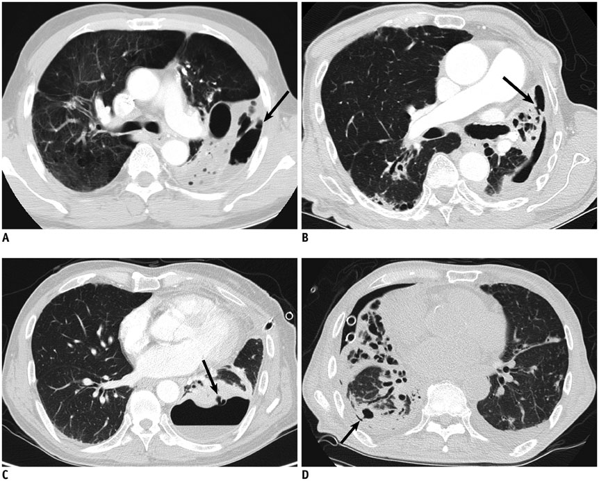
Fig. 2 CT images showing broncho-pleural fistula (arrows) in each case with hydropneumothorax associated with Mycobacterium avium complex lung disease (A–D, Cases 1–3, and 5, respectively).
Reference
-
1. Daley CL, Griffith DE. Pulmonary non-tuberculous mycobacterial infections. Int J Tuberc Lung Dis. 2010; 14:665–671.2. Griffith DE, Aksamit T, Brown-Elliott BA, Catanzaro A, Daley C, Gordin F, et al. An official ATS/IDSA statement: diagnosis, treatment, and prevention of nontuberculous mycobacterial diseases. Am J Respir Crit Care Med. 2007; 175:367–416.3. Hoefsloot W, van Ingen J, Andrejak C, Angeby K, Bauriaud R, Bemer P, et al. The geographic diversity of nontuberculous mycobacteria isolated from pulmonary samples: an NTM-NET collaborative study. Eur Respir J. 2013; 42:1604–1613.4. Kendall BA, Winthrop KL. Update on the epidemiology of pulmonary nontuberculous mycobacterial infections. Semin Respir Crit Care Med. 2013; 34:87–94.5. Koh WJ, Chang B, Jeong BH, Jeon K, Kim SY, Lee NY, et al. Increasing recovery of nontuberculous mycobacteria from respiratory specimens over a 10-year period in a tertiary referral hospital in South Korea. Tuberc Respir Dis (Seoul). 2013; 75:199–204.6. Kwon YS, Koh WJ. Diagnosis of pulmonary tuberculosis and nontuberculous mycobacterial lung disease in Korea. Tuberc Respir Dis (Seoul). 2014; 77:1–5.7. Aksamit TR. Mycobacterium avium complex pulmonary disease in patients with pre-existing lung disease. Clin Chest Med. 2002; 23:643–653.8. Chung MJ, Lee KS, Koh WJ, Lee JH, Kim TS, Kwon OJ, et al. Thin-section CT findings of nontuberculous mycobacterial pulmonary diseases: comparison between Mycobacterium avium-intracellulare complex and Mycobacterium abscessus infection. J Korean Med Sci. 2005; 20:777–783.9. Lee G, Kim HS, Lee KS, Koh WJ, Jeon K, Jeong BH, et al. Serial CT findings of nodular bronchiectatic Mycobacterium avium complex pulmonary disease with antibiotic treatment. AJR Am J Roentgenol. 2013; 201:764–772.10. Lee G, Lee KS, Moon JW, Koh WJ, Jeong BH, Jeong YJ, et al. Nodular bronchiectatic Mycobacterium avium complex pulmonary disease. Natural course on serial computed tomographic scans. Ann Am Thorac Soc. 2013; 10:299–306.11. Koh WJ, Yu CM, Suh GY, Chung MP, Kim H, Kwon OJ, et al. Pulmonary TB and NTM lung disease: comparison of characteristics in patients with AFB smear-positive sputum. Int J Tuberc Lung Dis. 2006; 10:1001–1007.12. Jeon D. Tuberculous pleurisy: an update. Tuberc Respir Dis (Seoul). 2014; 76:153–159.13. Light RW. Update on tuberculous pleural effusion. Respirology. 2010; 15:451–458.14. Kim HJ, Lee HJ, Kwon SY, Yoon HI, Chung HS, Lee CT, et al. The prevalence of pulmonary parenchymal tuberculosis in patients with tuberculous pleuritis. Chest. 2006; 129:1253–1258.15. Ko JM, Park HJ, Kim CH. Pulmonary changes of pleural TB: up-to-date CT imaging. Chest. 2014; 146:1604–1611.16. Christensen EE, Dietz GW, Ahn CH, Chapman JS, Murry RC, Anderson J, et al. Initial roentgenographic manifestations of pulmonary Mycobacterium tuberculosis, M kansasii, and M intracellularis infections. Chest. 1981; 80:132–136.17. Lynch DA, Simone PM, Fox MA, Bucher BL, Heinig MJ. CT features of pulmonary Mycobacterium avium complex infection. J Comput Assist Tomogr. 1995; 19:353–360.18. Nagaia T, Akiyama M, Mita Y, Tomizawa T, Dobashi K, Mori M. Mycobacterium avium complex pleuritis accompanied by diabetes mellitus. Diabetes Res Clin Pract. 2000; 48:99–104.19. Okada Y, Ichinose Y, Yamaguchi K, Kanazawa M, Yamasawa F, Kawashiro T. Mycobacterium avium-intracellulare pleuritis with massive pleural effusion. Eur Respir J. 1995; 8:1428–1429.20. Park SU, Koh WJ, Kwon OJ, Park HY, Jun HJ, Joo EJ, et al. Acute pneumonia and empyema caused by Mycobacterium intracellulare. Intern Med. 2006; 45:1007–1010.21. Yanagihara K, Tomono K, Sawai T, Miyazaki Y, Hirakata Y, Kadota J, et al. Mycobacterium avium complex pleuritis. Respiration. 2002; 69:547–549.22. Jeong BH, Jeon K, Park HY, Kim SY, Lee KS, Huh HJ, et al. Intermittent antibiotic therapy for nodular bronchiectatic Mycobacterium avium complex lung disease. Am J Respir Crit Care Med. 2015; 191:96–103.23. Koh WJ, Jeong BH, Jeon K, Lee NY, Lee KS, Woo SY, et al. Clinical significance of the differentiation between Mycobacterium avium and Mycobacterium intracellulare in M avium complex lung disease. Chest. 2012; 142:1482–1488.24. Koh WJ, Jeong BH, Jeon K, Lee SY, Shin SJ. Therapeutic drug monitoring in the treatment of Mycobacterium avium complex lung disease. Am J Respir Crit Care Med. 2012; 186:797–802.25. Hansell DM, Bankier AA, MacMahon H, McLoud TC, Müller NL, Remy J. Fleischner Society: glossary of terms for thoracic imaging. Radiology. 2008; 246:697–722.26. Stern EJ, Sun H, Haramati LB. Peripheral bronchopleural fistulas: CT imaging features. AJR Am J Roentgenol. 1996; 167:117–120.27. Ferrer J. Pleural tuberculosis. Eur Respir J. 1997; 10:942–947.28. Kakugawa T, Mukae H, Kajiki S, Tanaka A, Yamayoshi T, Inoue M, et al. Mycobacterium avium pleuritis in a non-immunocompromised patient. Intern Med. 2008; 47:1727–1731.29. Kotani K, Hirose Y, Endo S, Yamamoto H, Makihara S. Surgical treatment of atypical Mycobacterium intracellulare infection with chronic empyema: a case report. J Thorac Cardiovasc Surg. 2005; 130:907–908.30. Kim HY, Song KS, Goo JM, Lee JS, Lee KS, Lim TH. Thoracic sequelae and complications of tuberculosis. Radiographics. 2001; 21:839–858. discussion 859-860.31. Lois M, Noppen M. Bronchopleural fistulas: an overview of the problem with special focus on endoscopic management. Chest. 2005; 128:3955–3965.32. Kim SJ, Park J, Lee H, Lee YJ, Park JS, Cho YJ, et al. Risk factors for deterioration of nodular bronchiectatic Mycobacterium avium complex lung disease. Int J Tuberc Lung Dis. 2014; 18:730–736.33. Shu CC, Lee LN, Wang JT, Chien YJ, Wang JY, Yu CJ. Taiwan Anti-Mycobacteria Investigation (TAMI) Group. Non-tuberculous mycobacterial pleurisy: an 8-year single-centre experience in Taiwan. Int J Tuberc Lung Dis. 2010; 14:635–641. 4 p following 641.34. Kawamoto H, Yamagata M, Nakashima H, Kambe M, Okamoto N, Yamane K, et al. Development of a case of Mycobacterium avium complex disease from right pleural effusion. Nihon Kokyuki Gakkai Zasshi. 2000; 38:706–709.35. Koh WJ, Ko Y, Kim CK, Park KS, Lee NY. Rapid diagnosis of tuberculosis and multidrug resistance using a MGIT 960 system. Ann Lab Med. 2012; 32:264–269.
- Full Text Links
-
- Actions
-
Cited
- CITED
-
- Close
- Share
-
- Similar articles
-
- Mycobacterium intracellulare pulmonary infection accompanied with pleural effusion
- Pleural Calcification as a Manifestation of Paragonimiasis: A Report of Two Cases
- The Management of Delayed Post-Pneumonectomy Broncho-Pleural Fistula and Esophago-Pleural Fistula
- A Case of Mycobacterium avium Pulmonary Disease with Massive Pleural Effusion in an HIV-negative, Nonimmunosuppressed Patient: Using PCR-Restriction Fragment Length Polymorphism Assay
- Mycobacterium intracellulare Pleurisy Identified on Liquid Cultures of the Pleural Fluid and Pleural Biopsy
