Corneal Thickness Measurements Using 2 Kinds of Spectral Domain Optical Coherence Tomography, Pentacam, Ultrasound Pachymetry
- Affiliations
-
- 1The Institute of Ophthalmology and Optometry, Department of Ophthalmology, Ewha Womans University School of Medicine, Seoul, Korea. jrmoph@ewha.ac.kr
- KMID: 2355406
- DOI: http://doi.org/10.3341/jkos.2016.57.10.1527
Abstract
- PURPOSE
To compare the measurements of central corneal thickness (CCT) obtained using two kinds of spectral domain optical coherence tomography (OCT), Pentacam®, and ultrasound pachymetry (USP).
METHODS
CCT was measured by Cirrus OCT®, Spectralis OCT®, Pentacam®, and USP in 32 eyes from 32 subjects without ocular disease of the anterior segment.
RESULTS
The average CCT measurements using Cirrus OCT®, Spectralis OCT®, Pentacam®, and USP were 549.2 ± 28.7 µm, 545.2 ± 25.4 µm, 554.0 ± 27.8 µm, and 548.4 ± 27.9 µm respectively. The measurements were significantly highly correlated with each other (Pearson's correlation coefficient r > 0.9, all p-values < 0.001), but were significantly different (p < 0.001). The CCT 95% limits of agreement between Cirrus OCT® and Spectralis OCT®, Cirrus OCT® and Pentacam®, Cirrus OCT® and USP, Spectralis OCT® and Pentacam®, and Spectralis OCT® and USP were 27.70 µm, 26.1 µm, 26.97 µm, 22.91 µm, 35.59 µm, and 32.15 µm, respectively.
CONCLUSIONS
The CCT values measured using the four devices were highly correlated with each other, but the measurement using Pentacam® was significantly thicker than that using USP. The measurements of the two kinds of spectral domain OCT were similar to those using USP. Therefore, these differences should be considered in clinical use, and measurements cannot be considered interchangeable.
Figure
-
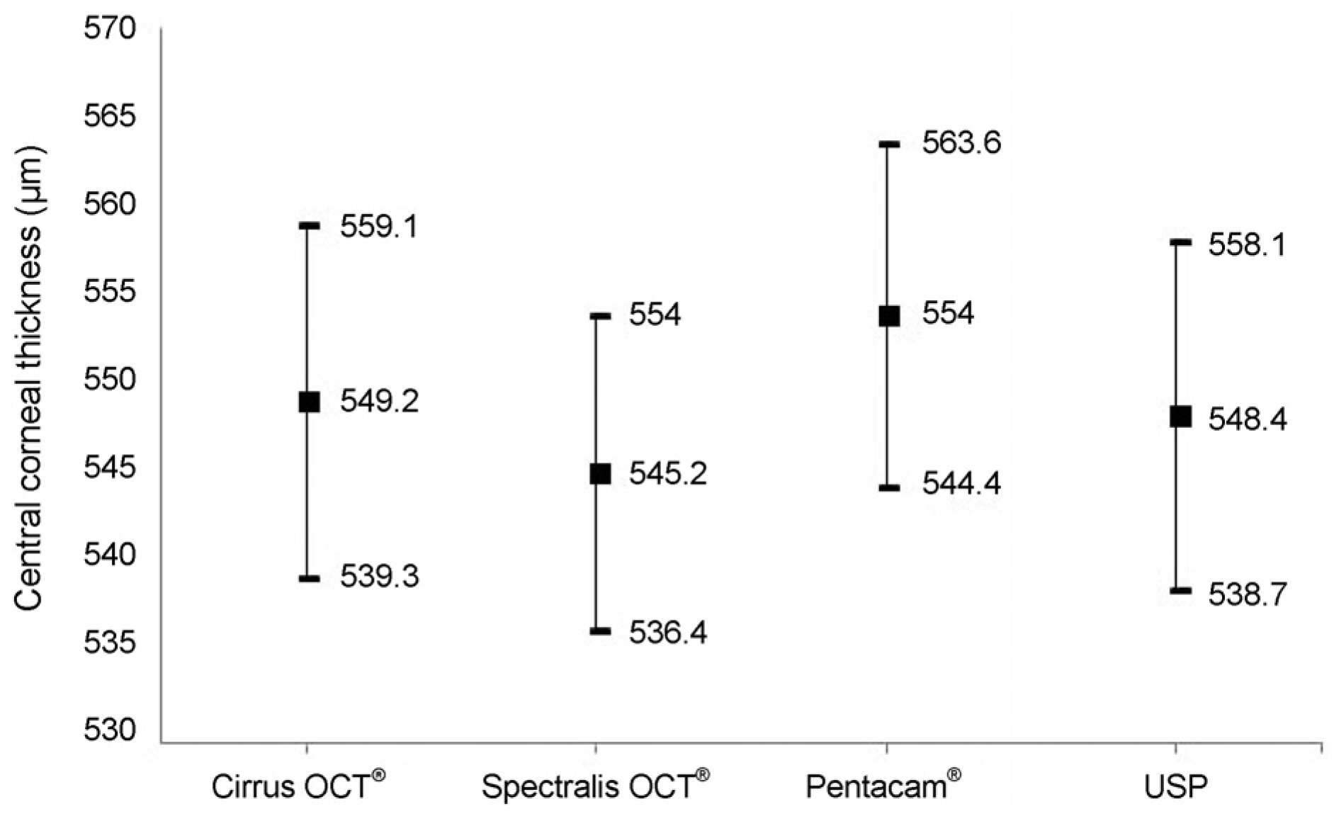
Figure 1. Mean and 95% confidence interval of central corneal thickness by Cirrus OCT®, Spectralis OCT®, Pentacam®, USP. OCT = optical coherence tomography; USP = ultrasound pachymetry.
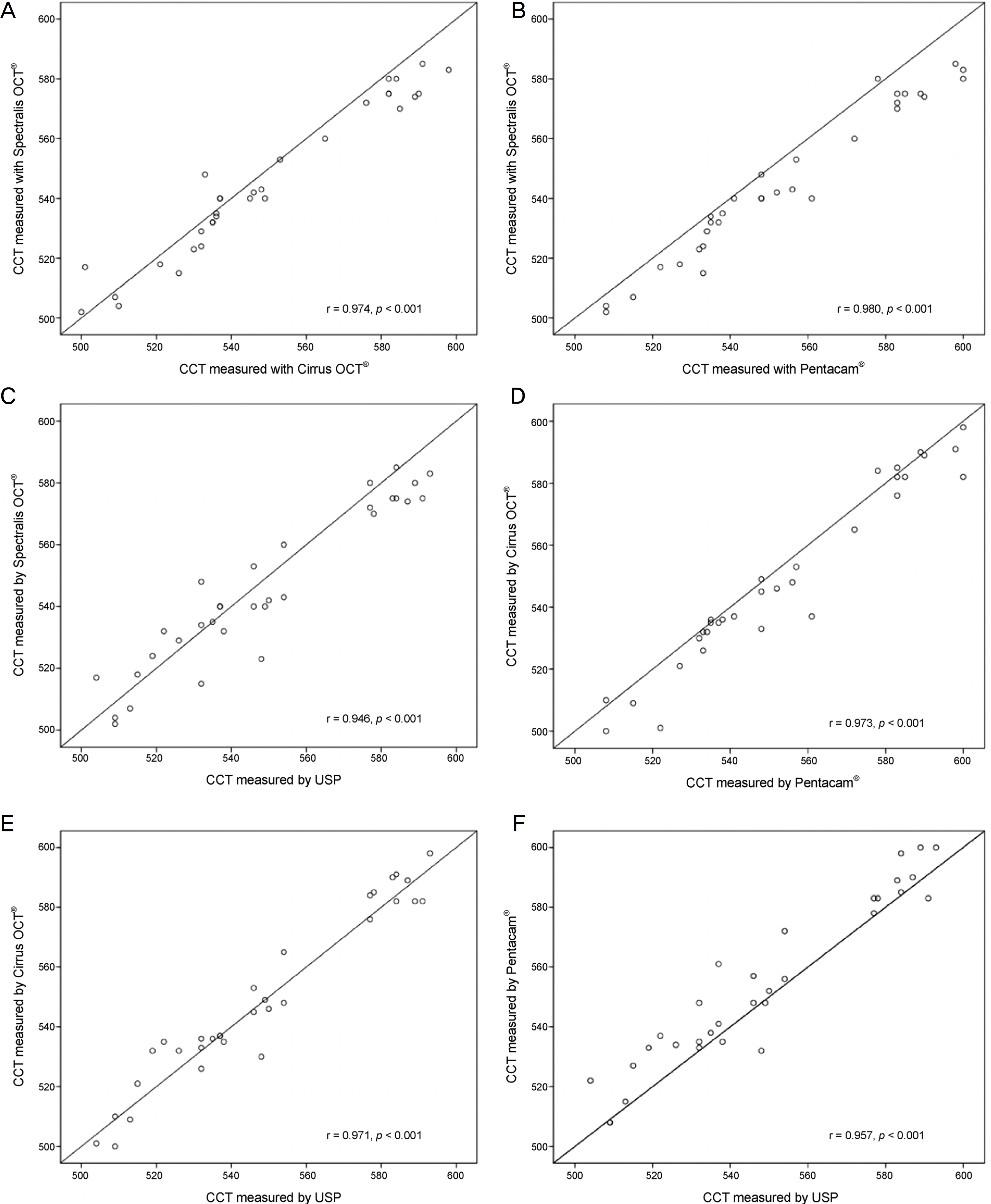
Figure 2. Scatter plots of central corneal thickness (μ m) between 4 methods. (A) Spectralis OCT® and Cirrus OCT®, (B) Spectralis OCT® and Pentacam®, (C) Spectralis OCT® and USP, (D) Cirrus OCT® and Pentacam®, (E) Cirrus OCT® and USP, (F) Pentacam® and USP. CCT = central corneal thickness; OCT = optical coherence tomography; USP = ultrasound pachymetry.
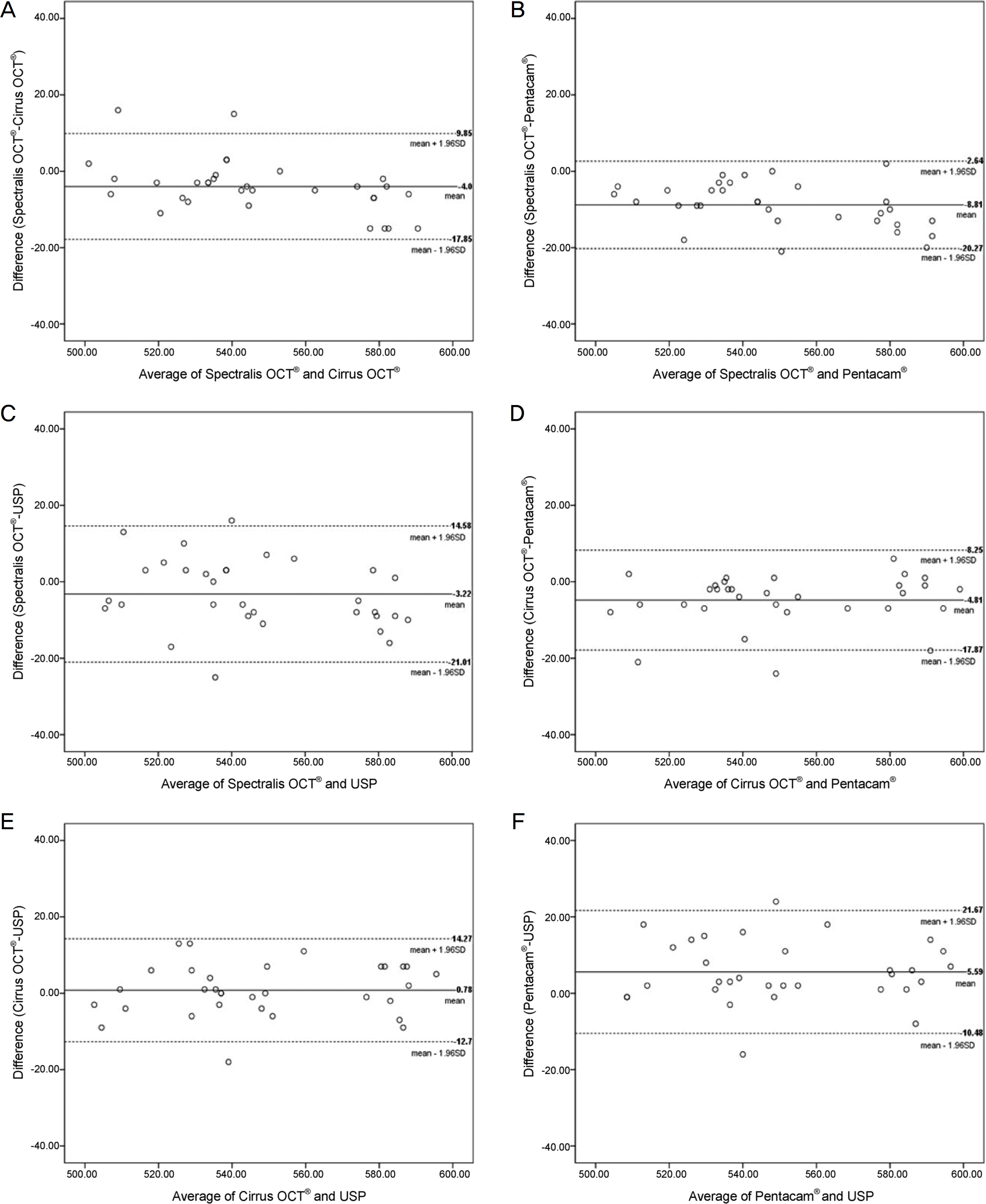
Figure 3. Bland-Altman plot of central corneal thickness (μ m) between 4 methods. (A) Spectralis OCT® and Cirrus OCT®, (B) Spectralis OCT® and Pentacam®, (C) Spectralis OCT® and USP, (D) Cirrus OCT® and Pentacam®, (E) Cirrus OCT® and USP, (F) Pentacam® and USP. OCT = optical coherence tomography; USP = ultrasound pachymetry; SD = standard deviation.
Reference
-
References
1. Faucher A, Grégoire J, Blondeau P. Accuracy of Goldmann abdominal after refractive surgery. J Cataract Refract Surg. 1997; 23:832–8.2. Fournier AV, Podtetenev M, Lemire J, et al. Intraocular pressure change measured by Goldmann tonometry after laser in situ keratomileusis. J Cataract Refract Surg. 1998; 24:905–10.
Article3. Chang DH, Stulting RD. Change in intraocular pressure abdominals after LASIK the effect of the refractive correction and the abdominal flap. Ophthalmology. 2005; 112:1009–16.4. Barkana Y, Gerber Y, Elbaz U, et al. Central corneal thickness measurement with the Pentacam Scheimpflug system, optical low-coherence reflectometry pachymeter, and ultrasound pachymetry. J Cataract Refract Surg. 2005; 31:1729–35.
Article5. Miranda MA, Radhakrishnan H, O'Donnell C. Repeatability of corneal thickness measured using an Oculus Pentacam. Optom Vis Sci. 2009; 86:266–72.
Article6. Solomon OD. Corneal indentation during ultrasonic pachometry. Cornea. 1999; 18:214–5.
Article7. Doughty MJ, Zaman ML. Human corneal thickness and its impact on intraocular pressure measures: a review and meta-analysis approach. Surv Ophthalmol. 2000; 44:367–408.8. Ling T, Ho A, Holden BA. Method of evaluating ultrasonic pachometers. Am J Optom Physiol Opt. 1986; 63:462–6.
Article9. Amano S, Honda N, Amano Y, et al. Comparison of central corneal thickness measurements by rotating Scheimpflug camera, ultrasonic pachymetry, and scanning-slit corneal topography. Ophthalmology. 2006; 113:937–41.
Article10. Leung DY, Lam DK, Yeung BY, Lam DS. Comparison between central corneal thickness measurements by ultrasound pachymetry and optical coherence tomography. Clin Exp Ophthalmol. 2006; 34:751–4.
Article11. Li H, Leung CK, Wong L, et al. Comparative study of central abdominal thickness measurement with abdominal optical coherence abdominal and visante optical coherence tomography. Ophthalmology. 2008; 115:796–801.e2.12. Rosa N, Lanza M, Borrelli M, et al. Comparison of central corneal thickness measured with Orbscan and Pentacam. J Refract Surg. 2007; 23:895–9.
Article13. Al-Mezaine HS, Al-Amro SA, Kangave D, et al. Comparison abdominal central corneal thickness measurements by oculus pentacam and ultrasonic pachymetry. Int Ophthalmol. 2008; 28:333–8.14. Bechmann M, Thiel MJ, Neubauer AS, et al. Central corneal abdominal measurement with a retinal optical coherence tomography device versus standard ultrasonic pachymetry. Cornea. 2001; 20:50–4.15. Fishman GR, Pons ME, Seedor JA, et al. Assessment of central corneal thickness using optical coherence tomography. J Cataract Refract Surg. 2005; 31:707–11.
Article16. Thomas J, Wang J, Rollins AM, Sturm J. Comparison of corneal thickness measured with optical coherence tomography, ultrasonic pachymetry, and a scanning slit method. J Refract Surg. 2006; 22:671–8.
Article17. Kim DW, Yi KY, Choi DG, Shin YJ. Corneal thickness measured by dual Scheimpflug, anterior segment optical coherence abdominal, and ultrasound pachymetry. J Korean Ophthalmol Soc. 2012; 53:1412–18.18. Yang YS, Koh JW. Utility of the noncontact specular microscopy for measurements of central corneal thickness. J Korean Ophthalmol Soc. 2014; 55:59–65.
Article19. Fu J, Wang X, Li S, et al. Comparative study of anterior segment measurement with Pentacam and anterior segment optical abdominal tomography. Can J Ophthalmol. 2010; 45:627–31.20. Azen SP, Burg KA, Smith RE, Maguen E. A comparison of three methods for the measurement of corneal thickness. Invest Ophthalmol Vis Sci. 1979; 18:535–8.
- Full Text Links
-
- Actions
-
Cited
- CITED
-
- Close
- Share
-
- Similar articles
-
- Utility of the Swept Source Optical Coherence Tomography for Measurements of Central Corneal Thickness
- Comparison of Corneal Thickness and Anterior Chamber Depth Measured With Orbscan, Pentacam, and Ultrasound Pachymetry
- Utility of the Anterior Segment Optical Coherence Tomography for Measurements of Central Corneal Thickness
- Reproducibility of IntraLASIK Flap Thickness Measured with Optical Coherence Tomography
- Corneal Thickness Measured by Dual Scheimpflug, Anterior Segment Optical Coherence Tomography, and Ultrasound Pachymetry
