Displaced Double-Layered Lateral Meniscus That Mimicked the Bucket-Handle Tear: a Case Report
- Affiliations
-
- 1Department of Radiology, Incheon St. Mary's Hospital, The Catholic University of Korea, Incheon, Korea. ssunky@catholic.ac.kr
- 2Department of Orthopedic Surgery, Incheon St. Mary's Hospital, The Catholic University of Korea, Incheon, Korea.
- 3Department of Pathology, Incheon St. Mary's Hospital, The Catholic University of Korea, Incheon, Korea.
- KMID: 2354801
- DOI: http://doi.org/10.13104/imri.2016.20.3.191
Abstract
- Among the various types of congenital meniscal anomalies, the double-layered lateral meniscus is extremely rare. The double-layered meniscus consists of both the upper additional and the lower normal meniscus. As the upper additional meniscus is mobile, it can be easily displaced, while the lower lateral meniscus is usually normal in shape and volume. A 42-year-old woman suffering from pain and locking of her left knee underwent Magnetic resonance imaging (MRI) examination and an arthroscopic surgery. A rare meniscal abnormality was seen in her left knee, which presented as a double-layered lateral meniscus with displacement. It was remarkable that the upper additional meniscus was displaced over the intercondylar eminence of the tibia and it mimicked a bucket-handle tear. Even though it is rare, it is necessary to consider the possibility of displaced double-layered meniscus in the differential diagnosis of a bucket-handle tear. Here, we report the MRI and arthroscopic findings of a displaced double-layered lateral meniscus, which was similar to the bucket-handle tear.
MeSH Terms
Figure
-
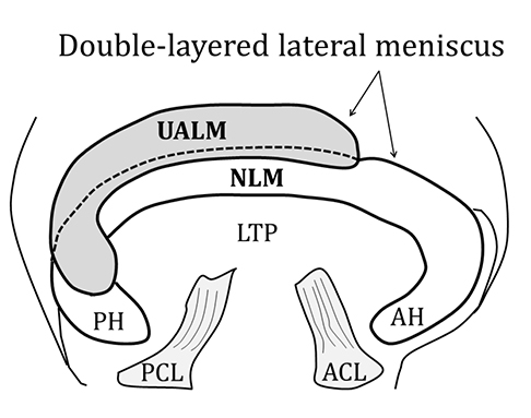
Fig. 1 A schematic illustration of the double layered lateral meniscus, which is characteristic that the upper additional meniscus overlays the normal lateral meniscus. ACL = anterior cruciate ligament, AH = anterior horn, LTP = lateral tibial plateau, NLM = normal lateral meniscus, PCL = posterior cruciate ligament, PH = posterior horn, UALM = upper additional lateral meniscus
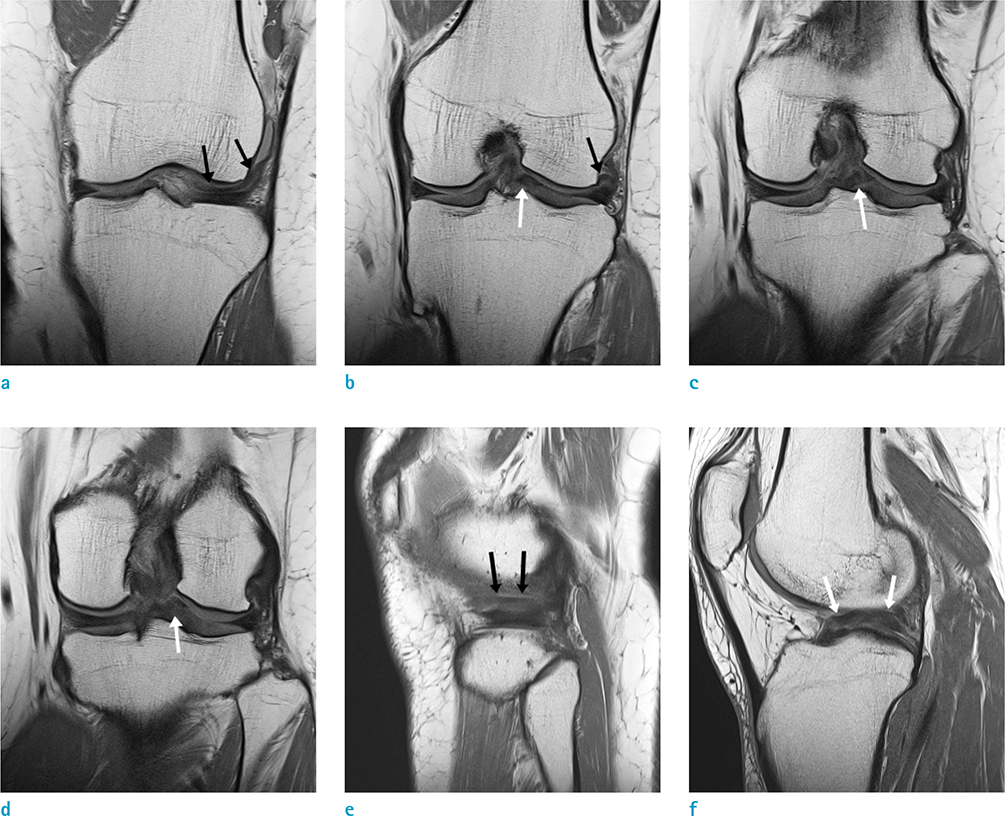
Fig. 2 Left knee MRI of a 42-year-old female presenting pain. A coronal proton density (PD) weighted spin-echo image (TR/TE, 2180/11) shows a double layered lateral meniscus, which includes the upper additional meniscus (black arrows) overlying the lower normal lateral meniscus (a, b) and a displaced meniscal fragment (white arrows) over the intercondylar eminence of tibia, mimicking the bucket-handle tear (b-d). A sagittal PD weighted spin-echo image (TR/TE, 2450/11) also shows the upper additional meniscus (black arrows) overlying the normal lateral meniscus (e) and a displaced upper additional meniscus (white arrows) over the intercondylar eminence of tibia (f).
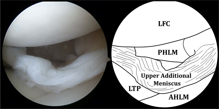
Fig. 3 Arthroscopic view of the lateral compartment shows that the upper additional meniscus overlying the normal lateral meniscus is displaced toward the intercondylar notch and mimics the bucket-handle tear; while the shape, margin and volume of the lower lateral meniscus is completely normal. These findings are suggesting a displaced double-layered meniscus. AHLM = anterior horn of lateral meniscus, LFC = lateral femoral condyle, LTP = lateral tibial plateau, PHLM = posterior horn of lateral meniscus
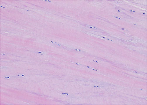
Fig. 4 Histology of the resected upper meniscus shows dense collagen fibers in multiple layers with normal organization, and fibro-chondrocytes that are linearly arranged, consistent with a typical meniscal tissue (H&E stain, × 200).
Reference
-
1. Suzuki S, Mita F, Ogishima H. Double-layered lateral meniscus: a newly found anomaly. Arthroscopy. 1991; 7:267–271.2. Lee KW, Yang DS, Choy WS. Dislocated double-layered lateral meniscus mimicking the bucket-handle tear. Orthopedics. 2013; 36:e1333–e1335.3. Dickhaut SC, DeLee JC. The discoid lateral-meniscus syndrome. J Bone Joint Surg Am. 1982; 64:1068–1073.4. Karahan M, Erol B. Accessory lateral meniscus: a case report. Am J Sports Med. 2004; 32:1973–1976.5. Saygi B, Yildirim Y, Senturk S, Sezgin Ramadan S, Gundes H. Accessory lateral discoid meniscus. Knee Surg Sports Traumatol Arthrosc. 2006; 14:1278–1280.6. Clark CR, Ogden JA. Development of the menisci of the human knee joint. Morphological changes and their potential role in childhood meniscal injury. J Bone Joint Surg Am. 1983; 65:538–547.7. Bailey WH, Blundell tge. An unusual abnormality affecting both knee joints in a child. Case report. J Bone Joint Surg Am. 1974; 56:814–816.8. Okahashi K, Sugimoto K, Iwai M, Oshima M, Fujisawa Y, Takakura Y. Double-layered lateral meniscus. J Orthop Sci. 2005; 10:661–664.9. Wang Q, Liu XM, Liu SB, Bai Y. Double-layered lateral meniscus. Knee Surg Sports Traumatol Arthrosc. 2011; 19:2050–2051.10. Takayama K, Kuroda R, Matsumoto T, et al. Bilateral double-layered lateral meniscus: a report of two cases. Knee Surg Sports Traumatol Arthrosc. 2009; 17:1336–1339.
- Full Text Links
-
- Actions
-
Cited
- CITED
-
- Close
- Share
-
- Similar articles
-
- Spontaneous Healing of a Displaced Bucket-Handle Tear of the Lateral Meniscus in a Child
- Ananalysis of the Clinical and MRI Findings of the Bucket: Handle Meniscal Tears of the Knee Joint
- Double-Layered Lateral Meniscus
- Arthroscopic Meniscectomy in Bucket Handle Tear of the Meniscus
- Double-Layered Lateral Meniscus: A Case Report
