Motor and Sensory Peripheral Neuropathy in a Patient Came after Acute Carbon Monoxide Intoxication: a Case Report with Magnetic Resonance Image
- Affiliations
-
- 1Department of Radiology, Dankook University Hospital, Cheonan, Korea. minipacs@daum.net
- KMID: 2354798
- DOI: http://doi.org/10.13104/imri.2016.20.3.175
Abstract
- Carbon monoxide (CO) intoxication is a leading cause of the variable neuropsychiatric impairment. Despite of widely known central nerve system complications after CO intoxication, peripheral neuropathy due to CO poisoning is rare and has been under-recognized. We report interesting case of a 29-year-old male who suffered from motor weakness and sensory abnormalities in his lower extremity following acute CO intoxication. The patient revealed direct and indirect signs of peripheral neuropathy of the left inferior gluteal and sciatic nerve on magnetic resonance imaging.
MeSH Terms
Figure
-

Fig. 1 On fluid-attenuated inversion recovery (FLAIR) images of follow-up brain MRI 4 weeks after CO intoxication (a, b), ill-defined high signal intensity lesions are seen along the both periventricular white matters, centrum semiovale (white arrows) and splenium of corpus callosum (arrowhead). On Diffusion-weighted image, focal high signal intensity is seen in the splenium of corpus callosum with corresponding low ADC map (arrowheads) (c, d), representing a b diffusion restriction.
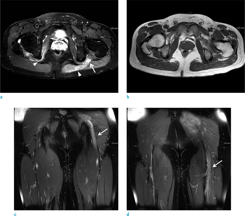
Fig. 2 On lower extremity MRI, axial fat-suppressed T2-weighted image (a) and T1-weighted image (b) show swollen left sciatic nerve with T2 hyperintensity (arrow). On fat-suppressed T2-weighted image (a), ill-defined high signal intensity lesions (arrowheads) are seen in left gluteus maximus and right obturator internus muscles, representing denervation edema. Two coronal fat-suppressed T2-weighted images (c, d) also reveal signal changes with swelling of left sciatic nerve at the levels of buttock and thigh (arrows).
Reference
-
1. Choi IS. Carbon monoxide poisoning: systemic manifestations and complications. J Korean Med Sci. 2001; 16:253–261.2. Choi IS. A clinical study of peripheral neuropathy in carbon monoxide intoxication. Yonsei Med J. 1982; 23:174–177.3. Weaver LK. Clinical practice. Carbon monoxide poisoning. N Engl J Med. 2009; 360:1217–1225.4. Zhang J, Piantadosi CA. Mitochondrial oxidative stress after carbon monoxide hypoxia in the rat brain. J Clin Invest. 1992; 90:1193–1199.5. Rahmani M, Belaidi H, Benabdeljlil M, et al. Bilateral brachial plexus injury following acute carbon monoxide poisoning. BMC Pharmacol Toxicol. 2013; 14:61.6. Jeong HJ, Kim CH, Kim MO, Jung HY. Bilateral femoral neuropathy in carbon monoxide poisoning: a case report. J Korean Assoc EMG Electrodiagn Med. 2011; 13:73–76.7. Jonas D, Conrad B, Von Einsiedel HG, Bischoff C. Correlation between quantitative EMG and muscle MRI in patients with axonal neuropathy. Muscle Nerve. 2000; 23:1265–1269.8. Andreisek G, Crook DW, Burg D, Marincek B, Weishaupt D. Peripheral neuropathies of the median, radial, and ulnar nerves: MR imaging features. Radiographics. 2006; 26:1267–1287.9. Wessig C, Koltzenburg M, Reiners K, Solymosi L, Bendszus M. Muscle magnetic resonance imaging of denervation and reinnervation: correlation with electrophysiology and histology. Exp Neurol. 2004; 185:254–261.
- Full Text Links
-
- Actions
-
Cited
- CITED
-
- Close
- Share
-
- Similar articles
-
- Motor Peripheral Neuropathy Involved Bilateral Lower Extremities Following Acute Carbon Monoxide Poisoning: A Case Report
- A Clinical Study of Peripheral Neuropathy in Carbon Monoxide Intoxication
- Multifocal Peripheral Neuropathies, Rhabdomyolysis, and Dermal Change in Carbon Monoxide Intoxication
- Delayed Anoxic Encephalopathy after Carbon Monoxide Poisoning: Evaluation of Therapeutic Effect by Serial Diffusion-Tensor Magnetic Resonance Imaging and Neurocognitive Test
- Acute Compartment Syndrome Which Causes Rhabdomyolysis by Carbon Monoxide Poisoning and Sciatic Nerve Injury Associated with It: A Case Report
