Ischial Tuberosity Avulsion Stress Fracture after Short Period of Repetitive Training
- Affiliations
-
- 1Department of Orthopedic Surgery, National Police Hospital, Seoul, Korea. ysr@police.go.kr
- KMID: 2354553
- DOI: http://doi.org/10.5371/hp.2016.28.3.187
Abstract
- Fatigue fracture of the pelvis is the form of fracture due to repetitive micro-stress accumulation, can be affected by a number of factors such as patient's nutritional status, biomechanics, social status and so on. Still there is no study about precise standard degree of external force that lead to stress fracture, but it may caused by compression force, traction force or complex force and others. Avulsion stress to ischial tuberosity or anterior superior iliac spine by attached muscle is known as the main factor for the avulsion fracture. This report will deal with 19 years old conscripted policeman who occurred ischial tuberosity avulsion fracture after training of 6-hour running for 5 days accompanying hip hyper-flexion motion. This reports aims to provide case study of stress fracture occurred after 5 days of exercise which is relatively short period who had no specific trauma history or pain.
Keyword
Figure
-
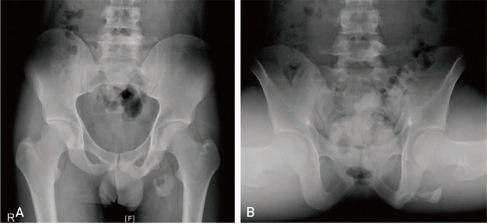
Fig. 1 (A) Initial pelvis anteropostrior view of the patient shows the avulsion fracture of the ischial tuberosity. (B) Pelvis frog leg view shows the bony fragment in different angle.
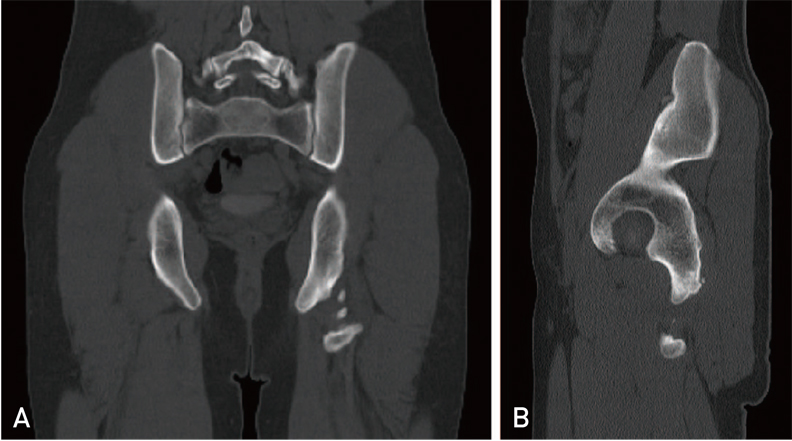
Fig. 2 (A) Coronal computed tomography image of pelvis shows the 35×40×20 mm sized bony fragment with multiple small fragments. (B) Sagittal computed tomography image of left pelvis shows the bony fragment, 15 mm distance from ischial tuberosity.
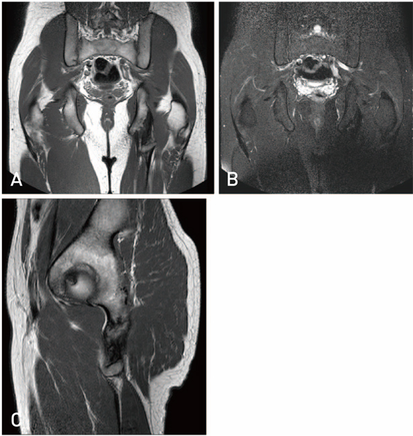
Fig. 3 (A) On the coronal T1-weighted image of pelvis, blurred margin of ischial tuberosity and distinct bony fragment suggest the avulsion stress fracture of the ischial tuberosity. Also, it shows the partial tear of the hamstring muscle origin area. (B) Coronal T2-weighted image of pelvis shows that there is no bone marrow edema at the ischial tuberosity. Then the fracture can be considered as an old fracture. (C) Magnetic resonance imaging T1-weighted sagittal view shows old avulsion fracture at ischial tuberosity which is mainly from conjoined origion of biceps femoris and semitendinosus tendons.
Reference
-
1. Kahanov L, Eberman LE, Games KE, Wasik M. Diagnosis, treatment, and rehabilitation of stress fractures in the lower extremity in runners. Open Access J Sports Med. 2015; 6:87–95.
Article2. Behrens SB, Deren ME, Matson A, Fadale PD, Monchik KO. Stress fractures of the pelvis and legs in athletes: a review. Sports Health. 2013; 5:165–174.
Article3. Gidwani S, Jagiello J, Bircher M. Avulsion fracture of the ischial tuberosity in adolescents--an easily missed diagnosis. BMJ. 2004; 329:99–100.
Article4. McKinney BI, Nelson C, Carrion W. Apophyseal avulsion fractures of the hip and pelvis. Orthopedics. 2009; 32:42.
Article5. Lenhart R, Thelen D, Heiderscheit B. Hip muscle loads during running at various step rates. J Orthop Sports Phys Ther. 2014; 44:766–774.
Article6. Schache AG, Dorn TW, Blanch PD, Brown NA, Pandy MG. Mechanics of the human hamstring muscles during sprinting. Med Sci Sports Exerc. 2012; 44:647–658.
Article7. Rockwood CA, Green DP, Court-Brown CM, et al. Rockwood and Green's fractures in adults. 8th ed. Philadelphia: Lippincott Williams & Wilkins;2015. p. 651–653.8. Lapp JM. Pelvic stress fracture: assessment and risk factors. J Manipulative Physiol Ther. 2000; 23:52–55.
Article
- Full Text Links
-
- Actions
-
Cited
- CITED
-
- Close
- Share
-
- Similar articles
-
- Sequela of Untreated Avulsion Fracutre of Ischial Tuberosity: Report of Two Cases
- Apophyseal Avulsion Fracture of Ischial Tuberosity during Soccer: A Case Report and Literature Review
- Avulsion Fracture of the Calcaneal Tuberosity: 2 Cases Report
- Isolated Avulsion Fracture of the Lesser Tuberosity of the Humerus: A Case Report
- Tibial Tuberosity Avulsion Fracture Combined with Meniscal Tear: A Case Report
