A pictorial review of signature patterns living in musculoskeletal ultrasonography
- Affiliations
-
- 1Department of Anesthesia and Pain Medicine, School of Medicine, Pusan National University, Yangsan, Korea. pain@pusan.ac.kr
- KMID: 2354214
- DOI: http://doi.org/10.3344/kjp.2016.29.4.217
Abstract
- The musculoskeletal system is mainly composed of the bones, muscles, tendons, and ligaments, in addition to nerves and blood vessels. The greatest difficulty in an ultrasonographic freeze-frame created by the examiner is recognition of the targeted structures without indicators, since an elephant's trunk may not be easily distinguished from its leg. It is not difficult to find descriptive ultrasonographic terms used for educational purposes, which help in distinguishing features of these structures either in a normal or abnormal anatomic condition. However, the terms sometimes create confusion when describing common objects, for example, in Western countries, pears have a triangular shape, but in Asia they are round. Skilled experts in musculoskeletal ultrasound have tried to express certain distinguishing features of anatomic landmarks using terms taken from everyday objects which may be reminiscent of that particular feature. This pictorial review introduces known signature patterns of distinguishing features in musculoskeletal ultrasound in a normal or abnormal condition, and may stir the beginners' interest to play a treasure-hunt game among unfamiliar images within a boundless ocean.
Keyword
MeSH Terms
Figure
-
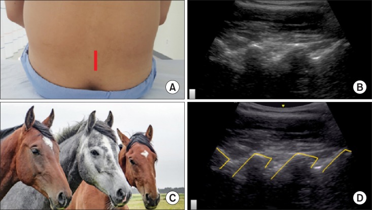
Fig. 1 A horse head sign of the lamina of the lumbar spine in a paramedian sagittal view. The lamina of the spine and its posterior acoustic shadow appear as a horse head in a paramedian sagittal view. (A) Probe location, (B) a paramedian sagittal ultrasonographic view, (C) a horse head, (D) a graphic overlay for (B).
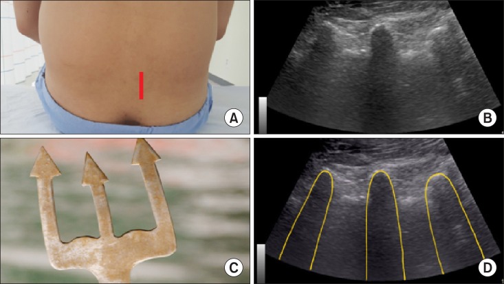
Fig. 2 A trident sign of the 3 consecutive transverse processes of the lumbar spine in a paramedian sagittal ultrasonographic view. The 3 consecutive transverse processes resemble as a trident, composed of hyperechoic curvilinear structures with finger-like acoustic shadowing beneath in a paramedian sagittal ultrasonographic view. (A) Probe location, (B) a paramedian sagittal ultrasonographic view, (C) a trident head, (D) a graphic overlay for (B).
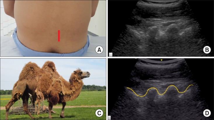
Fig. 3 A camel-hump sign of the lumbar articular process in a lumbar paramedian sagittal view. The superior and inferior articular processes resemble as a camel hump. (A) Probe location, (B) a paramedian sagittal ultrasonographic view, (C) a camel-hump, (D) a graphic overlay for (B).
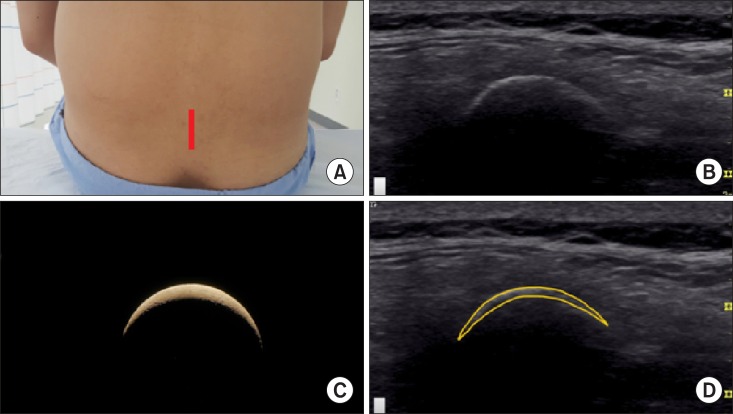
Fig. 4 A crescent sign of the lumbar midline dorsal spinous process in a median sagittal view. A lumbar midline dorsal spinous process with posterior acoustic shadowing appears as a crescent in a midline sagittal ultrasonographic view. (A) Probe location, (B) a midline sagittal ultrasonographic view, (C) a crescent, (D) a graphic overlay for (B).
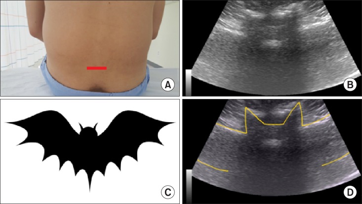
Fig. 5 A flying bat in a transverse midline interlaminar view. A flying bat in a midline transverse interlaminar view consists of both the transverse processes (wings) and the articular processes of the facet joints (ears) with the posterior (head; the ligament flavum and dura mater) and anterior (eyes; the dura mater, posterior longitudinal ligament, and vertebral body) complexes of the vertebral body. (A) Probe location, (B) a midline transverse ultrasonographic view, (C) a flying bat, (D) a graphic overlay for (B).
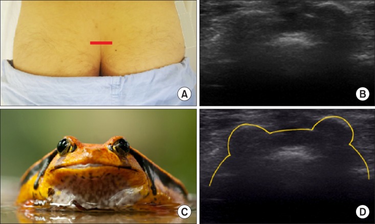
Fig. 6 Frog eyes sign of the 2 sacral cornua in a transverse view. The 2 sacral cornua above the sacral hiatus in a transverse ultrasonographic view appear as hyperechoic reversed U-shaped structures with acoustic shadowing, a frog eye sign, with the posterior surface of the sacrum. (A) Probe location, (B) a transverse ultrasonographic view, (C) a frog eye, (D) a graphic overlay for (B).
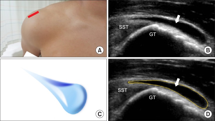
Fig. 7 A teardrop sign of the distended subacromial-subdeltoid bursa in a longitudinal view of the supraspinatus tendon. An abnormal subacromial-subdeltoid bursa with the most distended segment of the bursa most distal and most dependent may be shown as a teardrop in a transverse ultrasonographic view. (A) Probe location, (B) in a longitudinal view of the supraspinatus tendon, (C) a teardrop, (D) a graphic overlay for (B). Modified from Lew HL, Chen CP, Wang TG, Chew KT. Introduction to musculoskeletal diagnostic ultrasound: examination of the upper limb. Am J Phys Med Rehabil 2007; 86: 310-21.
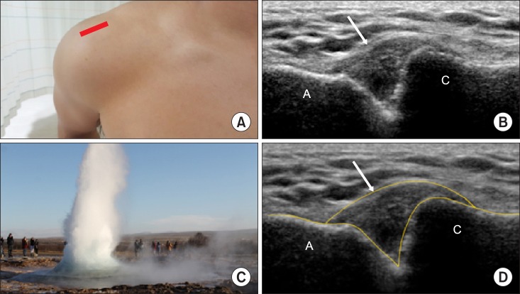
Fig. 8 A geyser sign of the cyst or ganglion projecting superiorly in a coronal across the acromioclavicular joint. It may present in rotator cuff tears. (A) Probe location, (B) a coronal across the acromioclavicular joint, (C) a teardrop, (D) a graphic overlay for (B). Modified from Beggs I. Shoulder ultrasound. Semin Ultrasound CT MR 2011; 32: 101-13.
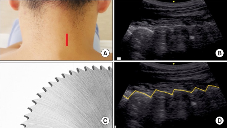
Fig. 9 A saw teeth sign of the cervical facet joints in a paramedian sagittal ultrasonographic view. The hyperechoic articular processes of the cervical facet joints in a sagittal ultrasonographic view resemble saw teeth in between the anechoic facet joint space. (A) Probe location, (B) a paramedian sagittal ultrasonographic view, (C) saw teeth, (D) a graphic overlay for (B).
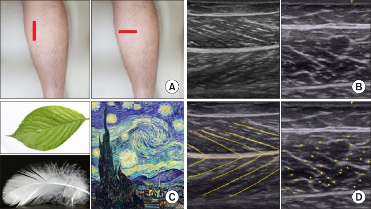
Fig. 10 A feather or veins on a leaf sign of the muscle in a longitudinal view and a starry night in a transverse ultrasonographic view in a medial gastrocnemius. The mixed echogenicity of muscle arises from the regular pattern of hypoechoic muscle fascicles within the hyperechoic perimysium and epimysium. (A) Probe location, (B) a longitudinal and transverse ultrasonographic view, (C) a feather or veins on a leaf and a starry night, (D) a graphic overlay for (B).
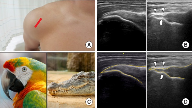
Fig. 11 A bird's (parrot's) beak in a normal supraspinous tendon attaching to the greater tubercle of the humerus and a crocodile's mouth in a ruptured supraspinous tendon coexisting with a cortical irregularity on the greater tubercle in a longitudinal view. (A) Probe location, (B) a longitudinal ultrasonographic view, (C) a bird's (parrot's) beak and a crocodile's mouth (D) graphic overlays for (B).
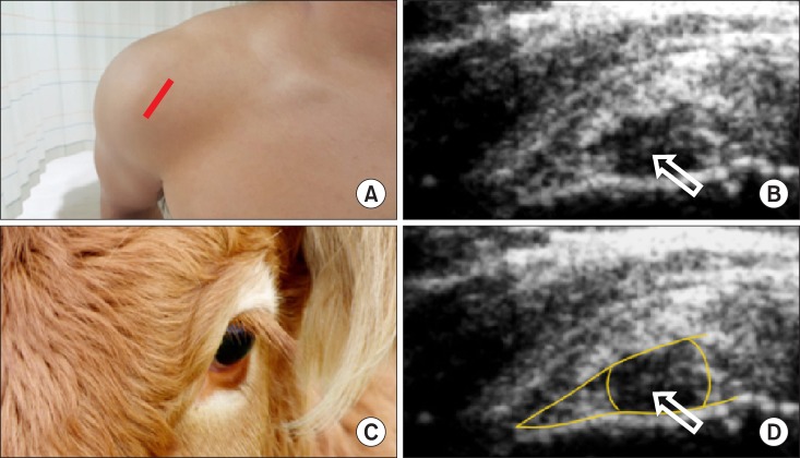
Fig. 12 A bull's sign of the rim rent tears of supraspinatus tendon in a transverse ultrasonographic view. Rim rent tears of the supraspinatus tendon, involving a small, articular surface avulsion adjacent tuberosity, appear as a small, hypoechoic defect with a central hyperechoic line on the articular surface. (A) Probe location, (B) a transverse ultrasonographic view, (C) a bull's sign, (D) a graphic overlay for (B). Modified from Lew HL, Chen CP, Wang TG, Chew KT. Introduction to musculoskeletal diagnostic ultrasound: examination of the upper limb. Am J Phys Med Rehabil 2007; 86: 310-21.
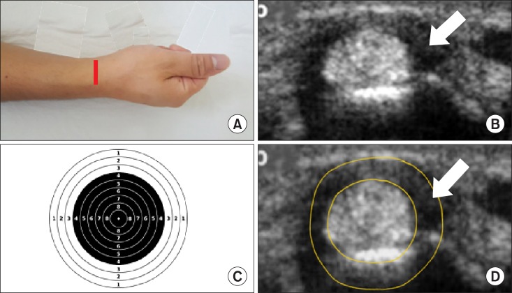
Fig. 13 A target sign of de Quervain tenosynovitis in a transverse ultrasonographic view. Peri-tendineal effusion in de Quervain tenosynovitis demonstrates a hypoechoic ring around the abductor pollicis longus and extensor pollicis brevis tendons. (A) Probe location, (B) a transverse ultrasonographic view, (C) a target sign, (D) a graphic overlay for (B). Modified from Lew HL, Chen CP, Wang TG, Chew KT. Introduction to musculoskeletal diagnostic ultrasound: examination of the upper limb. Am J Phys Med Rehabil 2007; 86: 310-21.
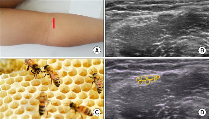
Fig. 14 A honeycomb sign of the peripheral nerve plexus in a transverse ultrasonographic view. (A) Probe location, (B) a transverse ultrasonographic view, (C) a honeycomb sign, (D) a graphic overlay for (B).
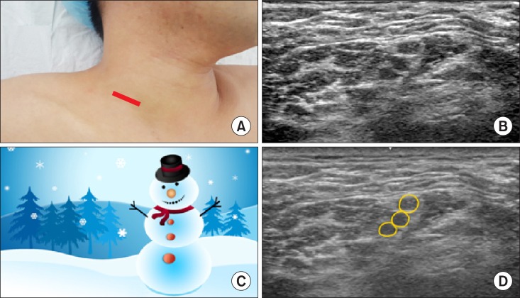
Fig. 15 A snowman sign of the consecutive nerve roots in a transverse ultrasonographic view. The C5, C6, and C7 nerve roots with an interscalene approach appear as a snowman. (A) Probe location, (B) a transverse ultrasonographic view, (C) a snowman sign, (D) a graphic overlay for (B).
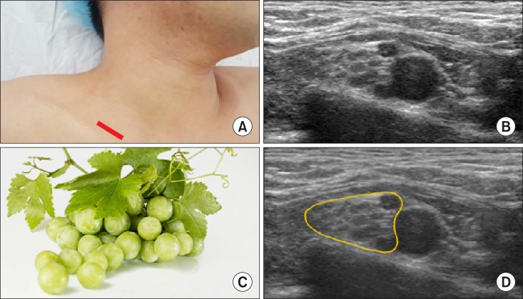
Fig. 16 A cluster of grapes sign of the subclavian and adjacent hyperechoic plexus in a transverse oblique view. (A) Probe location, (B) a transverse oblique view, (C) a cluster of grapes sign, (D) a graphic overlay for (B).
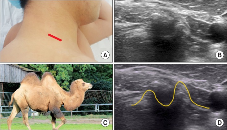
Fig. 17 A 2-humped camel sign of the bigger anterior and smaller posterior tubercle of the cervical transverse process in order to perform a selective nerve root block. (A) Probe location, (B) a transverse view, (C) a 2-humped camel sign, (D) a graphic overlay for (B).
Reference
-
1. Smith J, Finnoff JT. Diagnostic and interventional musculoskeletal ultrasound: part 2. Clinical applications. PM R. 2009; 1:162–177. PMID: 19627890.
Article2. Moore RE. 2010 musculoskeletal ultrasound for the extremities: a practical guide to sonography of the extremities. Scotts Valley (CA): Createspace;2010. p. 9–11.3. Karmakar MK, Li X, Kwok WH, Ho AM, Ngan Kee WD. Sonoanatomy relevant for ultrasound-guided central neuraxial blocks via the paramedian approach in the lumbar region. Br J Radiol. 2012; 85:e262–e269. PMID: 22010025.
Article4. Chin KJ, Karmakar MK, Peng P. Ultrasonography of the adult thoracic and lumbar spine for central neuraxial blockade. Anesthesiology. 2011; 114:1459–1485. PMID: 21422997.
Article5. Nomura JT, Leech SJ, Shenbagamurthi S, Sierzenski PR, O'Connor RE, Bollinger M, et al. A randomized controlled trial of ultrasound-assisted lumbar puncture. J Ultrasound Med. 2007; 26:1341–1348. PMID: 17901137.
Article6. Karmakar MK. Ultrasound-guided central neuraxial blocks. In : Narouze SN, editor. Atlas of ultrasound-guided procedures in interventional pain management. New York (NY): Springer;2011. p. 161–178.7. Möller I, Bong D, Naredo E, Filippucci E, Carrasco I, Moragues C, et al. Ultrasound in the study and monitoring of osteoarthritis. Osteoarthritis Cartilage. 2008; 16(Suppl 3):S4–S7. PMID: 18760636.
Article8. Razek AA, Fouda NS, Elmetwaley N, Elbogdady E. Sonography of the knee joint. J Ultrasound. 2009; 12:53–60. PMID: 23397073.
Article9. van Holsbeeck M, Strouse PJ. Sonography of the shoulder: evaluation of the subacromial-subdeltoid bursa. AJR Am J Roentgenol. 1993; 160:561–564. PMID: 8430553.
Article11. Narouze S, Peng PW. Ultrasound-guided interventional procedures in pain medicine: a review of anatomy, sonoanatomy, and procedures. Part II: axial structures. Reg Anesth Pain Med. 2010; 35:386–396. PMID: 20607896.
Article12. Woodhouse JB, McNally EG. Ultrasound of skeletal muscle injury: an update. Semin Ultrasound CT MR. 2011; 32:91–100. PMID: 21414545.
Article13. Tekin L, Kara M, Ozçakar L. When the parrot's beak becomes the crocodile's mouth: a story on shoulder ultrasound. Rheumatol Int. 2013; 33:2447–2448. PMID: 22811012.
Article14. Lew HL, Chen CP, Wang TG, Chew KT. Introduction to musculoskeletal diagnostic ultrasound: examination of the upper limb. Am J Phys Med Rehabil. 2007; 86:310–321. PMID: 17413545.15. van Geffen GJ, Moayeri N, Bruhn J, Scheffer GJ, Chan VW, Groen GJ. Correlation between ultrasound imaging, cross-sectional anatomy, and histology of the brachial plexus: a review. Reg Anesth Pain Med. 2009; 34:490–497. PMID: 19920425.
Article16. Kim YD, Yu JY, Shim J, Heo HJ, Kim H. Risk of encountering dorsal scapular and long thoracic nerves during ultrasound-guided interscalene brachial plexus block with nerve stimulator. Korean J Pain. 2016; 29:179–184. PMID: 27413483.
Article17. Lee MJ, Koo DJ, Choi YS, Lee KC, Kim HY. Dexamethasone or dexmedetomidine as local anesthetic adjuvants for ultrasound-guided axillary brachial plexus blocks with nerve stimulation. Korean J Pain. 2016; 29:29–33. PMID: 26839668.
Article18. Park SK, Sung MH, Suh HJ, Choi YS. Ultrasound guided low approach interscalene brachial plexus block for upper limb surgery. Korean J Pain. 2016; 29:18–22. PMID: 26839666.
Article19. Kim YH. Ultrasound phantoms to protect patients from novices. Korean J Pain. 2016; 29:73–77. PMID: 27103961.
Article
- Full Text Links
-
- Actions
-
Cited
- CITED
-
- Close
- Share
-
- Similar articles
-
- RE: Musculoskeletal Applications of Elastography: a Pictorial Essay of Our Initial Experience
- Musculoskeletal Applications of Elastography: a Pictorial Essay of Our Initial Experience
- Value of Ultrasound in Rheumatologic Diseases
- Sonographic Findings of Common Musculoskeletal Diseases in Patients with Diabetes Mellitus
- How to perform a functional assessment of the fetal heart: a pictorial review
