J Korean Ophthalmol Soc.
2016 Sep;57(9):1489-1492. 10.3341/jkos.2016.57.9.1489.
A Case of Hyphema after Selective Laser Trabeculoplasty
- Affiliations
-
- 1Department of Ophthalmology, Samsung Medical Center, Sungkyunkwan University School of Medicine, Seoul, Korea. ckee@skku.edu
- KMID: 2351884
- DOI: http://doi.org/10.3341/jkos.2016.57.9.1489
Abstract
- PURPOSE
To report a case of hyphema after selective laser trabeculoplasty (SLT) in a patient with pseudoexfoliative glaucoma.
CASE SUMMARY
A 77-year-old female was referred for elevation of intraocular pressure (IOP). Previously, she had been diagnosed with pseudoexfoliative glaucoma in the right eye and was using topical IOP-lowering agents. The best corrected visual acuity was 20/100 in the right eye and 20/40 in the left eye. IOP, measured with Goldmann applanation tonometer, was 32 mm Hg in the right eye and 20 mm Hg in the left eye. Gonioscopy revealed open-angle glaucoma with +2 trabecular meshwork pigmentation but without peripheral anterior synechiae or neovascularization. SLT was performed in the right eye. Two days later, the patient had sudden onset of blurred vision and pain in the right eye. Visual acuity was limited to light perception, and IOP was 34 mm Hg in the right eye. Slit-lamp examination revealed 1.1 mm hyphema with 4+ red blood cell count in the anterior chamber. Three weeks after the SLT, hyphema in the right eye disappeared, but IOP was measured to be 42 mm Hg. The patient underwent trabeculectomy in the right eye.
CONCLUSIONS
SLT is an effective means of lowering IOP with low risk of complications. However, hyphema can rarely occur after SLT and can affect the outcome of the treatment.
MeSH Terms
Figure
-
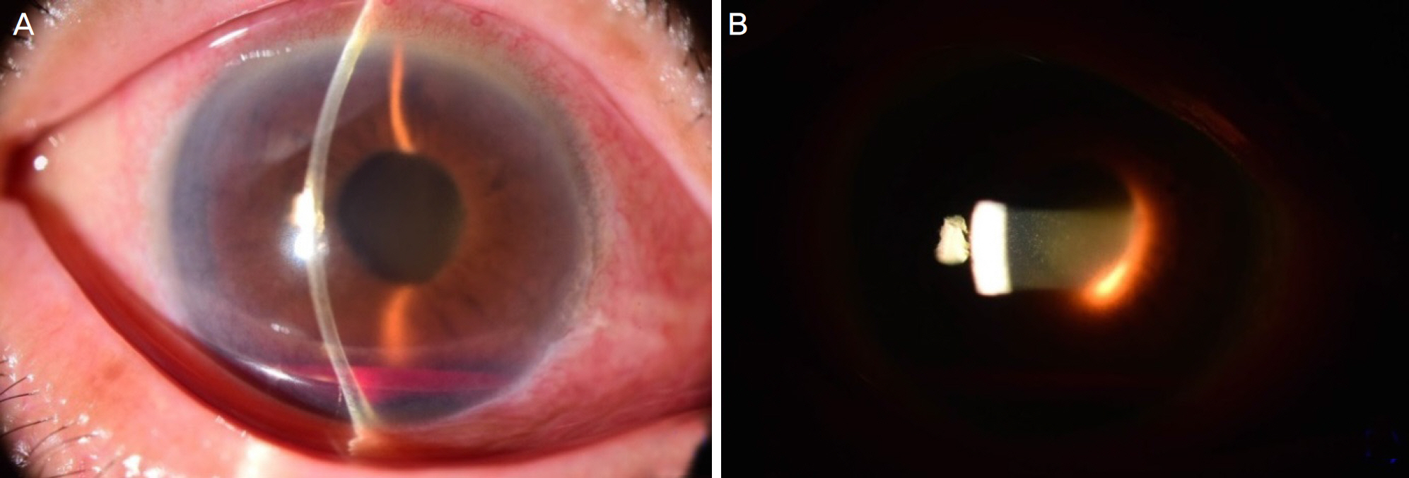
Figure 1. Anterior segment photographs, 2 days after selective laser trabeculoplasty. (A) Slit-lamp examination demonstrates hyphema of 1.1 mm in height. (B) 4+ red blood cell count is noted in the anterior chamber.
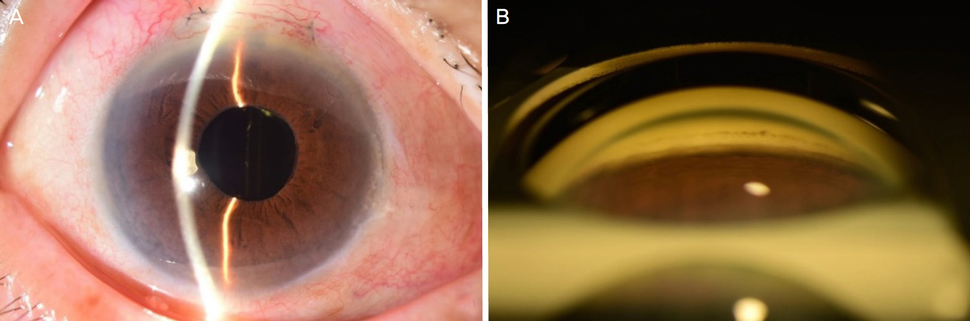
Figure 2. Anterior segment phorographs, 1 month after trabeculectomy. (A) Hyphema in the right eye has disappeared. (B) Gonioscopy revealed open-angle without any peripheral anterior synechiae or neovascularization.
Reference
-
References
1. Juzych MS, Chopra V, Banitt MR, et al. Comparison of long-term outcomes of selective laser trabeculoplasty versus argon laser abdominal in open-angle glaucoma. Ophthalmology. 2004; 111:1853–9.2. Latina MA, Sibayan SA, Shin DH, et al. Q-switched 532-nm Nd:YAG laser trabeculoplasty (selective laser trabeculoplasty): a multicenter, pilot, clinical study. Ophthalmology. 1998; 105:2082–8. discussion 2089–90.3. Martinez-de-la-Casa JM, Garcia-Feijoo J, Castillo A, et al. Selective vs argon laser trabeculoplasty: hypotensive efficacy, abdominal chamber inflammation, and postoperative pain. Eye (Lond). 2004; 18:498–502.4. Rubin L, Arnett J, Ritch R. Delayed hyphema after argon laser iridectomy. Ophthalmic Surg. 1984; 15:852–3.5. Kumar N, Feyi-Waboso A. Intractable secondary glaucoma from hyphema following YAG iridotomy. Can J Ophthalmol. 2005; 40:85–6.
Article6. Ritch R, Shields MB, Krupin T. The Glaucomas. 2nd ed.St. Louis: Mosby;1996. p. 1582.7. Kramer TR, Noecker RJ. Comparison of the morphologic changes after selective laser trabeculoplasty and argon laser trabeculoplasty in human eye bank eyes. Ophthalmology. 2001; 108:773–9.
Article8. Rhee DJ, Krad O, Pasquale LR. Hyphema following selective laser trabeculoplasty. Ophthalmic Surg Lasers Imaging. 2009; 40:493–4.
Article9. Shihadeh WA, Ritch R, Liebmann JM. Hyphema occurring during selective laser trabeculoplasty. Ophthalmic Surg Lasers Imaging. 2006; 37:432–3.
Article
- Full Text Links
-
- Actions
-
Cited
- CITED
-
- Close
- Share
-
- Similar articles
-
- Comparison of Short-term Outcomes of Argon Laser versus Selective Laser Trabeculoplasty in Open-Angle Glaucoma
- Comparison of Clinical Outcomes of Argon Laser Versus Selective Laser Trabeculoplasty in POAG
- Short-Term Clinical Outcomes of Laser Trabeculoplasty Using a 577-nm Wavelength Laser
- Clinical Result of Argon Laser Trabeculoplasty in Primary Open Angle Glaucoma
- Two-year Follow-up of Selective Laser Trabeculoplasty as Initial and Adjunctive Treatment for Ocular Hypertension and Open Angle Glaucoma
