J Korean Soc Radiol.
2010 Oct;63(4):371-378.
Visually Lossless Threshold: JPEG 2000 compression of Digital Chest Radiographs
- Affiliations
-
- 1Department of Radiology, Seoul National University College of Medicine, Seoul National University Hospital, Korea.
- 2Department of Radiation Applied Life Science, Seoul National University College of Medicine, Korea. kil210@snubhrad.snu.ac.kr
Abstract
- PURPOSE
To estimate the visual lossless threshold of Joint Photographic Experts Group (JPEG) 2000 compression digital chest radiograph images.
MATERIALS AND METHODS
Fifty (n=50) selected chest radiograph images were compressed to 5 different levels: reversible (as negative control) and irreversible 5:1, 10:1, 15:1, and 20:1. By alternately displaying the original image and its paired compressed image on the same monitor, five radiologists independently determined if the image pairs had detectable differences. For each reader, we compared the proportion of the image pairs (the compressed image and the original image) rated to have detectable differences between reversible compression and each of the four irreversible compressions using the exact test for paired proportions.
RESULTS
For each reader, the proportion of the image pairs rated to have detectable difference was not significantly different between the reversible and irreversible 5:1 and 10:1 compressions. However, the proportion significantly increased with 15:1 and 20:1 irreversible compressions, versus reversible compression in all readers (p=7.4x10⁻²² -0.027).
CONCLUSION
10:1 compressed chest radiograph images can be considered visually lossless and are therefore potentially acceptable for primary interpretation.
MeSH Terms
Figure
-
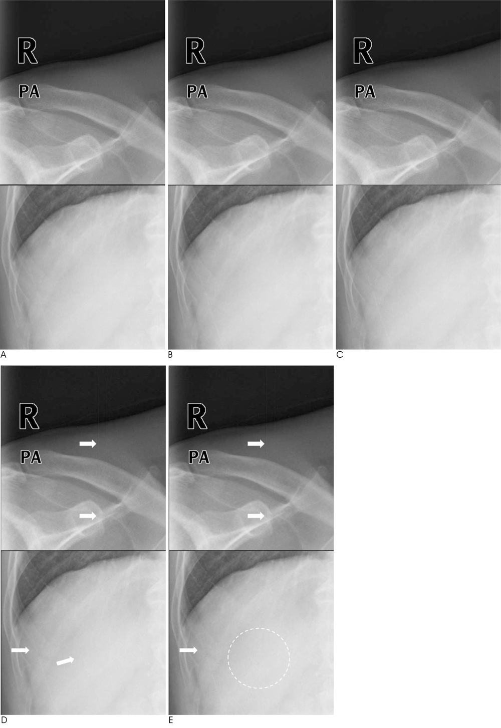
Fig. 1 JPEG 2000 compression artifacts in a chest radiograph image of a 59-year-old male with a mass in the right middle lung field. Irreversibly 5:1 (B) and 10:1 (C) compressed images are indistinguishable from the original (A). At a compression level of 15:1 (D), vertical linear artifact appears in the right part of the image (arrows). At a compression level of 20:1 (E), the manifestation of the artifacts is apparent (arrows) and the internal texture of the soft tissue is degraded (dashed circle). The compression artifacts are best demonstrated if the two images are alternately displayed on a single monochrome monitor calibrated according to the Digital Imaging and Communications in Medicine Gray Scale Standard Display Function.
Reference
-
1. Rubin GD. Data explosion: the challenge of multidetector-row CT. Eur J Radiol. 2000; 36:74–80.2. Rubin GD. 3-D imaging with MDCT. Eur J Radiol. 2003; 45:Suppl 1. S37–S41.3. Tamm EP, Thompson S, Venable SL, McEnery K. Impact of multislice CT on PACS resources. J Digit Imaging. 2002; 15:Suppl 1. 96–101.4. Lee KH, Lee HJ, Kim JH, Kang HS, Lee KW, Hong H, et al. Managing the CT data explosion: initial experiences of archiving volumetric datasets in a mini-PACS. J Digit Imaging. 2005; 18:188–195.5. Ko JP, Rusinek H, Naidich DP, McGuinness G, Rubinowitz AN, Leitman BS, et al. Wavelet compression of low-dose chest CT data: effect on lung nodule detection. Radiology. 2003; 228:70–75.6. Ko JP, Chang J, Bomsztyk E, Babb JS, Naidich DP, Rusinek H. Effect of CT image compression on computer-assisted lung nodule volume measurement. Radiology. 2005; 237:83–88.7. Ishigaki T, Sakuma S, Ikeda M, Itoh Y, Suzuki M, Iwai S. Clinical evaluation of irreversible image compression: analysis of chest imaging with computed radiography. Radiology. 1990; 175:739–743.8. Kido S, Ikezoe J, Kondoh H, Takeuchi N, Johkoh T, Kohno N, et al. Detection of subtle interstitial abnormalities of the lungs on digitized chest radiographs: acceptable data compression ratios. AJR Am J Roentgenol. 1996; 167:111–115.9. MacMahon H, Doi K, Sanada S, Montner SM, Giger ML, Metz CE, et al. Data compression: effect on diagnostic accuracy in digital chest radiography. Radiology. 1991; 178:175–179.10. Mori T, Nakata H. Irreversible data compression in chest imaging using computed radiography: an evaluation. J Thorac Imaging. 1994; 9:23–30.11. Ohgiya Y, Gokan T, Nobusawa H, Hirose M, Seino N, Fujisawa H, et al. Acute cerebral infarction: effect of JPEG compression on detection at CT. Radiology. 2003; 227:124–127.12. Slone RM, Foos DH, Whiting BR, Muka E, Rubin DA, Pilgram TK, et al. Assessment of visually lossless irreversible image compression: comparison of three methods by using an image-comparison workstation. Radiology. 2000; 215:543–553.13. Slone RM, Muka E, Pilgram TK. Irreversible JPEG compression of digital chest radiographs for primary interpretation: assessment of visually lossless threshold. Radiology. 2003; 228:425–429.14. Ringl H, Schernthaner RE, Kulinna-Cosentini C, Weber M, Schaefer-Prokop C, Herold CJ, et al. Lossy three-dimensional JPEG2000 compression of abdominal CT images: assessment of the visually lossless threshold and effect of compression ratio on image quality. Radiology. 2007; 245:467–474.15. Daly S. Application of a noise-adaptive contrast sensitivity function to image data compression. Opt Eng. 1990; 29:977–987.16. Aberle DR, Gleeson F, Sayre JW, Brown K, Batra P, Young DA, et al. The effect of irreversible image compression on diagnostic accuracy in thoracic imaging. Invest Radiol. 1993; 28:398–403.17. Savcenko V, Erickson BJ, Palisson PM, Persons KR, Manduca A, Hartman TE, et al. Detection of subtle abnormalities on chest radiographs after irreversible compression. Radiology. 1998; 206:609–616.18. Kalyanpur A, Neklesa VP, Taylor CR, Daftary AR, Brink JA. Evaluation of JPEG and wavelet compression of body CT images for direct digital teleradiologic transmission. Radiology. 2000; 217:772–779.19. Hansell DM, Bankier AA, MacMahon H, McLoud TC, Muller NL, Remy J. Fleischner Society: glossary of terms for thoracic imaging. Radiology. 2008; 246:697–722.20. Kim KJ, Kim B, Choi SW, Kim YH, Hahn S, Kim TJ, et al. Definition of compression ratio: difference between two commercial JPEG2000 program libraries. Telemed J E Health. 2008; 14:350–354.21. Lee KH, Kim YH, Kim BH, Kim KJ, Kim TJ, Kim HJ, et al. Irreversible JPEG 2000 compression of abdominal CT for primary interpretation: assessment of visually lossless threshold. Eur Radiol. 2007; 17:1529–1534.22. Woo HS, Kim KJ, Kim TJ, Hahn S, Kim B, Kim YH, et al. JPEG 2000 compression of abdominal CT: difference in tolerance between thin- and thick-section images. AJR Am J Roentgenol. 2007; 189:535–541.23. Kim KJ, Kim B, Lee KH, Kim TJ, Mantiuk R, Kang HS, et al. Regional difference in compression artifacts in low-dose chest CT images: effects of mathematical and perceptual factors. AJR Am J Roentgenol. 2008; 191:W30–W37.24. Kim B, Lee KH, Kim KJ, Mantiuk R, Kim HR, Kim YH. Artifacts in slab average-intensity-projection images reformatted from JPEG 2000 compressed thin-section abdominal CT data sets. AJR Am J Roentgenol. 2008; 190:W342–W350.25. Kim B, Lee KH, Kim KJ, Mantiuk R, Hahn S, Kim TJ, et al. Prediction of perceptible artifacts in JPEG 2000-compressed chest CT images using mathematical and perceptual quality metrics. AJR Am J Roentgenol. 2008; 190:328–334.26. Kim TJ, Lee KH, Kim B, Kim KJ, Chun EJ, Bajpai V, et al. Regional variance of visually lossless threshold in compressed chest CT images: lung versus mediastinum and chest wall. Eur J Radiol. 2009; 69:483–488.27. Newcombe RG. Two-sided confidence intervals for the single proportion: comparison of seven methods. Stat Med. 1998; 17:857–872.28. Liddell FD. Simplified exact analysis of case-referent studies: matched pairs; dichotomous exposure. J Epidemiol Community Health. 1983; 37:82–84.29. Fleiss JL, Cuzick J. The reliability of dichotomous judgements: unequal numbers of judges per subjects. Appl Psychol Meas. 1979; 3:537–542.30. Erickson BJ, Manduca A, Palisson P, Persons KR, Earnest F 4th, Savcenko V, et al. Wavelet compression of medical images. Radiology. 1998; 206:599–607.
- Full Text Links
-
- Actions
-
Cited
- CITED
-
- Close
- Share
-
- Similar articles
-
- Comparison of JPEG and wavelet compression on intraoral digital radiographic images
- Comparison of Compression Methods of Radiological Images for the Personal Archive
- 3-D Lossless Volumetric Medical Image Compression Using 3-D Integer Wavelet Transform and Lifting Steps
- Evaluation of compression ratios using JPEG 2000 on diagnostic images in dentistry
- Influence of Image Compression on the Interpretation of Optical Coherence Tomography in Diabetic Macular Edema
