Influence of Image Compression on the Interpretation of Optical Coherence Tomography in Diabetic Macular Edema
- Affiliations
-
- 1Department of Ophthalmology, Kim's Eye Hospital, Konyang University College of Medicine, Seoul, Korea. kjh7997@daum.net
- KMID: 2217115
- DOI: http://doi.org/10.3341/jkos.2014.55.9.1320
Abstract
- PURPOSE
To evaluate the influence of image compression on optical coherence tomography (OCT) images in eyes with diabetic macular edema (DME).
METHODS
Twenty eyes of 30 patients diagnosed with DME were included in this retrospective observational case series. Horizontal OCT scans centered at the center of the fovea were conducted using spectral-domain OCT (Spectral OCT/SLO(R)). The images were exported to Tag Image File Format (TIFF) and then transformed to 10, 5, and 1 quality of Joint Photographic Experts Group (JPEG) format using Photoshop. OCT images were taken before and after intravitreal bevacizumab injection. The presence of intraretinal fluid, foveolar detachment, and photoreceptor inner segment/outer segment (IS/OS) disruption were evaluated in each image.
RESULTS
The mean (+/- standard deviation) size of TIFF images and 10, 5 and 1 quality JPEG images were 1712.0, 183.3 +/- 6.8, 90.9 +/- 4.3, 42.8 +/- 1.4 kilobytes (KB), respectively, before the injection and 1712.0, 189.5 +/- 9.1, 94.9 +/- 5.6, 43.4 +/- 1.8 KB, respectively, after the injection. The presence of intraretinal fluid, foveolar detachment, and photoreceptor IS/OS disruption identified in TIFF images was also identified in the compressed JPEG images.
CONCLUSIONS
Quality of retinal OCT image did not influence the estimation of DME despite the JPEG image being compressed to approximately 1/40 of the original TIFF image size.
MeSH Terms
Figure
-
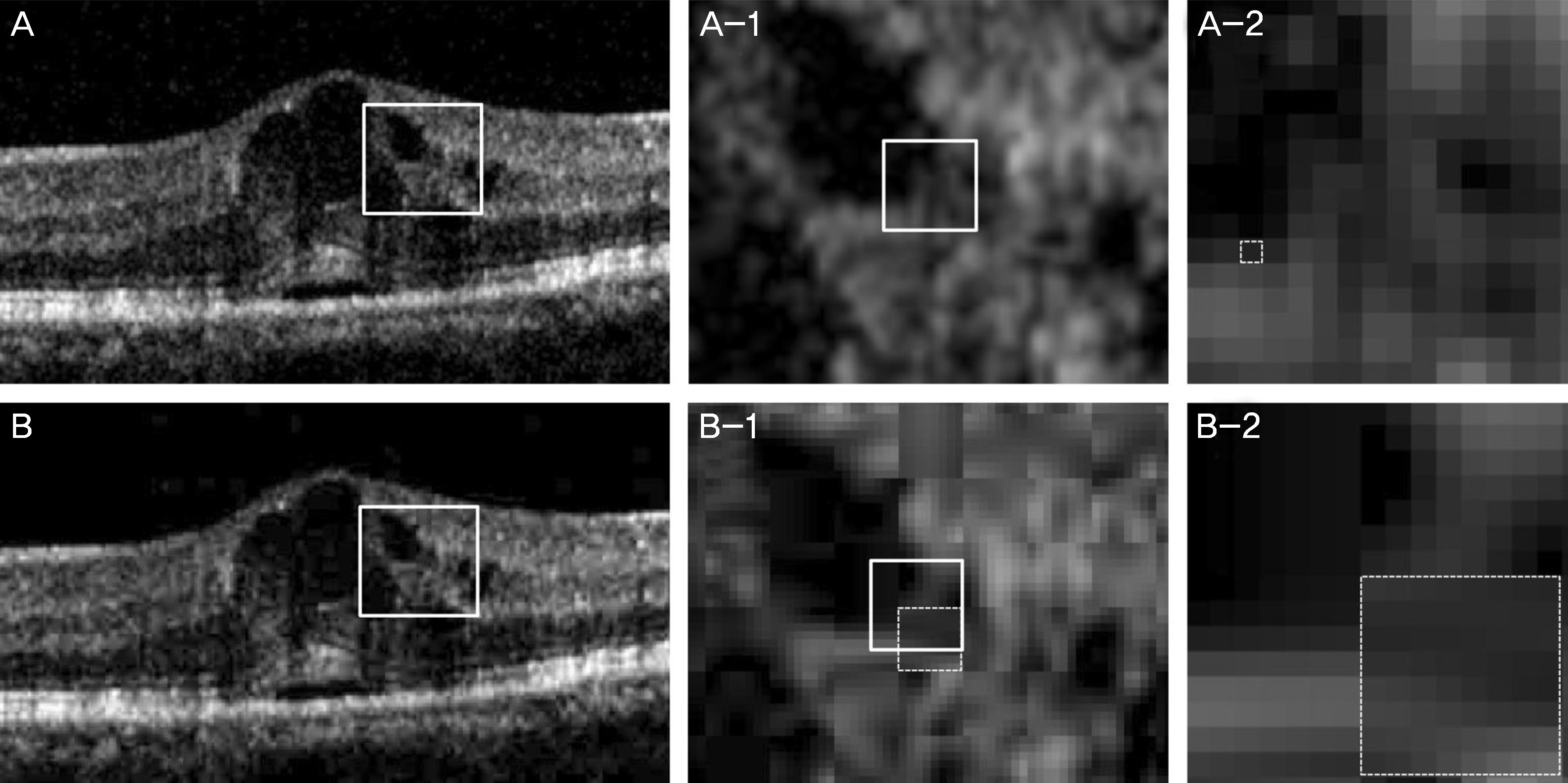
Figure 1. Comparison of image quality between an image of Tag Image File Format (TIFF) format (A, A-1, A-2) and images of 1 quality of Joint Photographic Experts Group (JPEG) compression format (B, B-1, B-2) in an eye with diabetic macular edema before treatment. A-1, B-1: ×600% magnified view of areas enclosed by white square (A, B), A-2, B-1: ×3,200% magnified view of areas enclosed by white square (A-1, B-1). Magnified views show the distorted image. The image was composed of small squares (dotted square, A-2) in the TIFF image, whereas the basic unit composing the JPEG image was relative large square (dotted square, B-1, B-2) which composed of 8×8 small squares. However, the presence of subfoveal serous detachment and small intraretinal cysts were clearly identified even in compressed image.
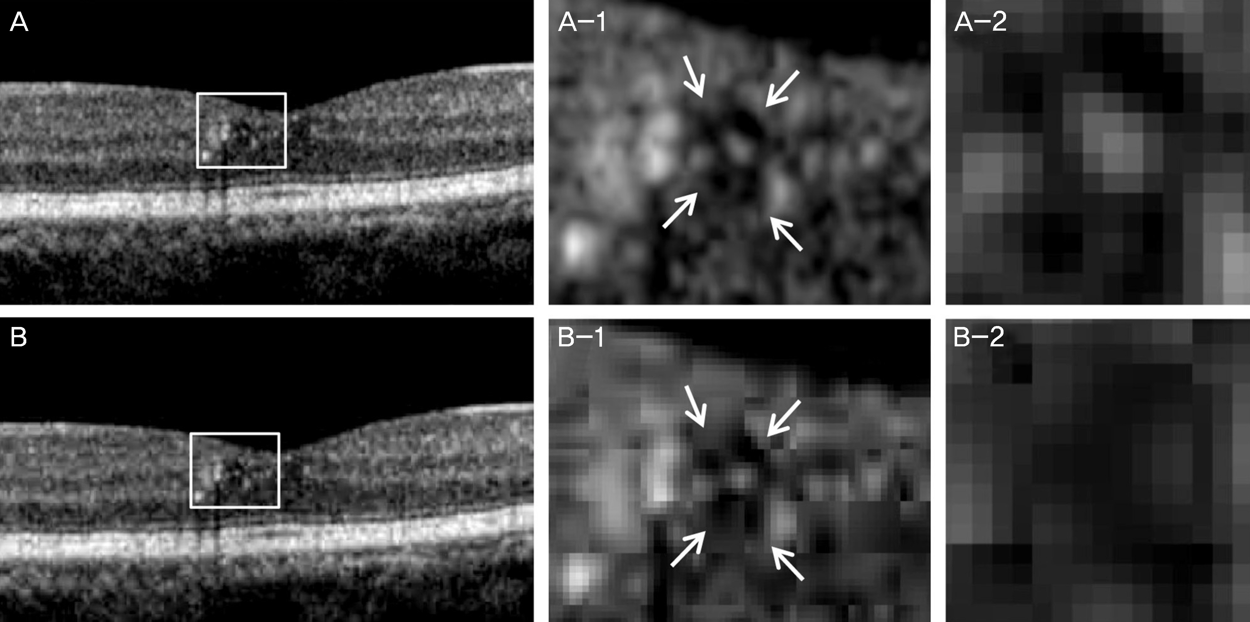
Figure 2. Comparison of image quality between an image of Tag Image File Format (TIFF) format (A, A-1, A-2) and images of 1 quality of Joint Photographic Experts Group (JPEG) compression format (B, B-1, B-2) in an eye with diabetic macular edema after treatment. A-1, B-1: ×600% magnified view of areas enclosed by white square (A, B), A-2, B-2: ×3,200% magnified view of areas indicated by white arrows (A-1, B-1). Magnified view shows the distorted pixels in compressed JPEG image (B-1, B-2). The presence of very small intraretinal cysts white arrows was clearly identified even in compressed image (B-1).
Reference
-
References
1. Alam S, Zawadzki RJ, Choi S, et al. Clinical application of rapid serial fourier-domain optical coherence tomography for macular imaging. Ophthalmology. 2006; 113:1425–31.
Article2. Nassif N, Cense B, Park BH, et al. In vivo human retinal imaging by ultrahigh-speed spectral domain optical coherence tomography. Opt Lett. 2004; 29:480–2.
Article3. Lee SH, Chung H, Kim HC. Subfoveal choroidal thickness in fellow eyes of patients with central serous chorioretinopathy. J Korean Ophthalmol Soc. 2012; 53:982–7.
Article4. Kim JH, Kang SW, Park DY, et al. Asymmetric elongation of foveal tissue after macular hole surgery and its impact on metamorphopsia. Ophthalmology. 2012; 119:2133–40.
Article5. Brown DM, Chen E, Mariani A, Major JC Jr. Super-dose anti-VEGF (SAVE) trial: 2.0 mg intravitreal ranibizumab for recalcitrant neovascular macular degeneration-primary end point. Ophthalmology. 2013; 120:349–54.
Article6. Cohen SY, Dubois L, Nghiem-Buffet S, et al. Spectral domain optical coherence tomography analysis of macular changes in tilted disk syndrome. Retina. 2013; 33:1338–45.
Article7. Yeung L, Lima VC, Garcia P, et al. Correlation between spectral domain optical coherence tomography findings and fluorescein angiography patterns in diabetic macular edema. Ophthalmology. 2009; 116:1158–67.
Article8. Bolz M, Kriechbaum K, Simader C, et al. In vivo retinal morphology after grid laser treatment in diabetic macular edema. Ophthalmology. 2010; 117:538–44.
Article9. Haritoglou C, Kook D, Neubauer A, et al. Intravitreal bevacizumab (Avastin) therapy for persistent diffuse diabetic macular edema. Retina. 2006; 26:999–1005.
Article10. Paccola L, Costa RA, Folgosa MS, et al. Intravitreal triamcinolone versus bevacizumab for treatment of refractory diabetic macular oedema (IBEME study). Br J Ophthalmol. 2008; 92:76–80.
Article11. Yanyali A, Aytug B, Horozoglu F, Nohutcu AF. Bevacizumab (Avastin) for diabetic macular edema in previously vitrectomized eyes. Am J Ophthalmol. 2007; 144:124–6.
Article12. Jeong JH, Kim ES, Lee JK, et al. The effects of intravitreal bevacizumab injection according to the type of diabetic macular edema. J Korean Ophthalmol Soc. 2010; 51:700–6.
Article13. Lee SJ, Kim ET, Moon YS. Intravitreal bevacizumab alone versus combined with macular photocoagulation in diabetic macular edema. Korean J Ophthalmol. 2011; 25:299–304.
Article14. Maheshwary AS, Oster SF, Yuson RM, et al. The association between percent disruption of the photoreceptor inner segment-outer segment junction and visual acuity in diabetic macular edema. Am J Ophthalmol. 2010; 150:63–7.e1.
Article15. Otani T, Yamaguchi Y, Kishi S. Correlation between visual acuity and foveal microstructural changes in diabetic macular edema. Retina. 2010; 30:774–80.
Article16. Shin HJ, Lee SH, Chung H, Kim HC. Association between photoreceptor integrity and visual outcome in diabetic macular edema. Graefes Arch Clin Exp Ophthalmol. 2012; 250:61–70.
Article17. Sakamoto A, Nishijima K, Kita M, et al. Association between foveal photoreceptor status and visual acuity after resolution of diabetic macular edema by pars plana vitrectomy. Graefes Arch Clin Exp Ophthalmol. 2009; 247:1325–30.
Article18. Lim JH, Kim IH, Bae GH, et al. Analysis of optical coherence tomographic patterns and clinical courses in diabetic macular edema after treatment. J Korean Ophthalmol Soc. 2014; 55:222–9.
Article19. Kim JH, Kang SW, Ha HS, et al. Overestimation of subfoveal choroidal thickness by measurement based on horizontally compressed optical coherence tomography images. Graefes Arch Clin Exp Ophthalmol. 2013; 251:1091–6.
Article
- Full Text Links
-
- Actions
-
Cited
- CITED
-
- Close
- Share
-
- Similar articles
-
- The Correlation between Visual Acuity and Patterns of Diabetic Macular Edema in OCT Images
- Risk Factors for Diffuse Diabetic Macular Edema as Classified by Optical Coherence Tomography
- Diurnal Variation of Macular Thickness in Diabetic Macular Edema
- Quantitative Analysis of Diabetic Macular Edema by Optical Coherence Tomography
- A Case of Macular Edema after Rosiglitazone Use
