Ann Dermatol.
2016 Aug;28(4):495-496. 10.5021/ad.2016.28.4.495.
Fibro-Osseous Pseudotumor of the Digit: A Diagnostic Pitfall of Extraskeletal Osteosarcoma
- Affiliations
-
- 1Department of Dermatology, VHS Medical Center, Seoul, Korea. choikohy@gmail.com
- KMID: 2344822
- DOI: http://doi.org/10.5021/ad.2016.28.4.495
Abstract
- No abstract available.
MeSH Terms
Figure
-
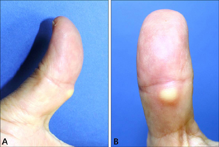
Fig. 1 Painful, skin-colored, solitary mass on the left thumb. (A) Lateral view, (B) palmar side view.
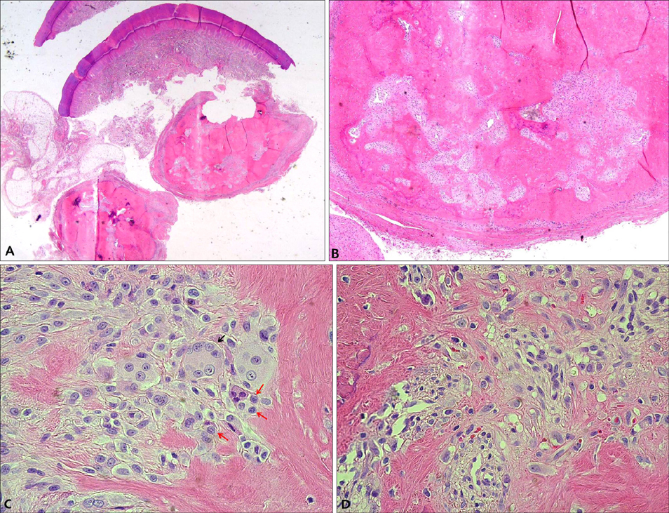
Fig. 2 (A) Scanning view demonstrating the subcutaneous trabecular mass (H&E, ×1), (B) osteoid formation surrounded by fibrotic band with trabecular margin and calcified foci (H&E, ×40), (C) multi-nucleated osteoclast (black arrow) and a few of mitotic cells (red arrows; H&E, ×400), (D) biphasic immature pattern of spindle cellular portion and bony portion, and cells having an irregular size and atypical nucleus (H&E, ×400).
Reference
-
1. Chaudhry IH, Kazakov DV, Michal M, Mentzel T, Luzar B, Calonje E. Fibro-osseous pseudotumor of the digit: a clinicopathological study of 17 cases. J Cutan Pathol. 2010; 37:323–329.
Article2. Lee SS, Baker BL, Gapp JD, Rosenberg AE, Googe PB. Ossifying plexiform tumor. J Cutan Pathol. 2015; 42:61–65.
Article3. Lee EJ, Lee JH, Shin MK, Lee SW, Haw CR. Acral angioosteoma cutis. Ann Dermatol. 2011; 23:Suppl 1. S105–S107.
Article4. Nishio J, Iwasaki H, Soejima O, Naito M, Kikuchi M. Rapidly growing fibro-osseous pseudotumor of the digits mimicking extraskeletal osteosarcoma. J Orthop Sci. 2002; 7:410–413.
Article5. Sleater J, Mullins D, Chun K, Hendricks J. Fibro-osseous pseudotumor of the digit: a comparison to myositis ossificans by light microscopy and immunohistochemical methods. J Cutan Pathol. 1996; 23:373–377.
Article
- Full Text Links
-
- Actions
-
Cited
- CITED
-
- Close
- Share
-
- Similar articles
-
- A Case of Recurrent Fibro-osseous Pseudotumor of the Digit
- A Case of Fibro-osseous Pseudotumor of the Digit
- Fibro-osseous Pseudotumor of the Digits: A case report
- A Case of Rapidly Growing Fibro-osseous Pseudotumor of the Toe after Trauma
- Radiological Features of a Fibro-Osseous Pseudotumor in the Digit: A Case Report
