Radiological Features of a Fibro-Osseous Pseudotumor in the Digit: A Case Report
- Affiliations
-
- 1Department of Radiology, Incheon St. Mary's Hospital, The Catholic University of Korea College of Medicine, Incheon, Korea. ssunky@catholic.ac.kr
- 2Department of Orthopedic Surgery, Incheon St. Mary's Hospital, The Catholic University of Korea College of Medicine, Incheon, Korea.
- 3Department of Pathology, Incheon St. Mary's Hospital, The Catholic University of Korea College of Medicine, Incheon, Korea.
- KMID: 1964266
- DOI: http://doi.org/10.3348/jksr.2015.73.2.131
Abstract
- A fibro-osseous pseudotumor is a rare ossifying soft tissue lesion, which is thought to be a reactive rather than a neoplastic lesion, developing due to repeated trauma. The lesion mostly occurs in the subcutaneous tissue of the proximal phalanx of the digit and predominantly affects young adults, with a slight predominance in females. The clinicopathological features can mimic those of malignant soft tissue lesions, and diagnosis can be difficult. Less is known about the radiological appearance of the lesion, including magnetic resonance imaging (MRI) features, than about histological signs. Here, we report radiological findings, including MRI features, of a fibro-osseous pseudotumor of the digit in a young female.
MeSH Terms
Figure
-
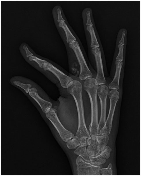
Fig. 1 A plain radiograph demonstrating a fairly circumscribed, ossified soft tissue mass, adjacent to, but separate from, bone, overlying the proximal phalanx of the right middle finger, without any periosteal reaction or cortical change.

Fig. 2 Hand MRI of a 19-year-old female presenting palpable mass on the digit. A. An axial T1-weighted spin-echo image (TR/TE, 912/13) showing a soft tissue mass (arrows) overlying the proximal phalanx of the right middle finger, without any periosteal or cortical reaction. The peripheral component of the mass is of intermediate signal intensity, and an internal region is hyperintense, likely indicating osseous differentiation surrounded by fibroblastic proliferation. B. A coronal T2-weighted spin-echo image (TR/TE, 4360/80) showing an ovoid-shaped hyperintense mass (arrows), with several foci of intermediate signal intensities, overlying the proximal phalanx of the right middle finger, suggesting mixed osseous and fibroblastic proliferation. C. On the axial fat-suppressed contrast-enhanced T1-weighted image (TR/TE, 650/12), the peripheral component of intermediate signal intensity on the axial T1-weighted spin-echo image demonstrates contrast enhancement (arrows), suggesting a fibroblastic proliferative area, whereas the central area (exhibiting little contrast enhancement) is thought to be the osseous portion. TE = echo time, TR = repetition time

Fig. 3 Photomicrographs of the fibro-osseous pseudotumor. A. The tumor features a peripheral fasciitis-like stroma of fibroblasts and a central irregular immature osteoid (H&E, × 40). B. There is mild to moderate cellular proliferation of fibroblasts at the periphery, and immature osteoid formation with osteoblastic rimming (H&E, × 100). H&E = hematoxylin and eosin
Reference
-
1. Javdan M, Tahririan MA. Fibro-osseous pseudotumor of the digit. Adv Biomed Res. 2012; 1:31.2. Tan KB, Tan SH, Aw DC, Lee YS. Fibro-osseous pseudotumor of the digit: presentation as an enlarging erythematous cutaneous nodule. Dermatol Online J. 2010; 16:7.3. Sleater J, Mullins D, Chun K, Hendricks J. Fibro-osseous pseudotumor of the digit: a comparison to myositis ossificans by light microscopy and immunohistochemical methods. J Cutan Pathol. 1996; 23:373–377.4. Hashmi AA, Faridi N, Edhi MM, Jafri A, Khan M. Fibro-osseous pseudotumor of the digit presenting as an ulcerated lesion: a case report. Int Arch Med. 2014; 7:4.5. Moosavi CA, Al-Nahar LA, Murphey MD, Fanburg-Smith JC. Fibroosseous [corrected] pseudotumor of the digit: a clinicopathologic study of 43 new cases. Ann Diagn Pathol. 2008; 12:21–28.6. Calisir C, Kocman AE, Oztunali C, Arik D, Uzuner M, Cetin C. Imaging findings of an extradigital fibro-osseous pseudotumor. Jpn J Radiol. 2014; 32:613–617.7. Nalbantoglu U, Gereli A, Kocaoglu B, Aktas S. Fibro-osseous pseudotumor of the digits: a rare tumor in an unusual location. J Hand Surg Am. 2008; 33:273–276.8. Dupree WB, Enzinger FM. Fibro-osseous pseudotumor of the digits. Cancer. 1986; 58:2103–2109.9. Chaudhry IH, Kazakov DV, Michal M, Mentzel T, Luzar B, Calonje E. Fibro-osseous pseudotumor of the digit: a clinicopathological study of 17 cases. J Cutan Pathol. 2010; 37:323–329.10. Kransdorf MJ, Meis JM. From the archives of the AFIP. Extraskeletal osseous and cartilaginous tumors of the extremities. Radiographics. 1993; 13:853–884.
