Treatment of Serous Retinal Detachment Associated with Choroidal Ischemia with Intravitreal Bevacizumab Following Brain Surgery
- Affiliations
-
- 1Institute of Vision Research, Department of Ophthalmology, Yonsei University College of Medicine, Seoul, Korea. minkim76@yuhs.ac
- KMID: 2344268
- DOI: http://doi.org/10.3341/kjo.2014.28.5.424
Abstract
- No abstract available.
MeSH Terms
-
Angiogenesis Inhibitors/*therapeutic use
Bevacizumab/*therapeutic use
Choroid/*blood supply
Ciliary Arteries/pathology
Fluorescein Angiography
Humans
Intravitreal Injections
Ischemia/*drug therapy/etiology/physiopathology
Male
Meningeal Neoplasms/surgery
Meningioma/surgery
Neurosurgical Procedures/*adverse effects
Retinal Detachment/*drug therapy/etiology/physiopathology
Subretinal Fluid
Vascular Endothelial Growth Factor A/antagonists & inhibitors
Visual Acuity/physiology
Young Adult
Angiogenesis Inhibitors
Bevacizumab
Vascular Endothelial Growth Factor A
Figure
-
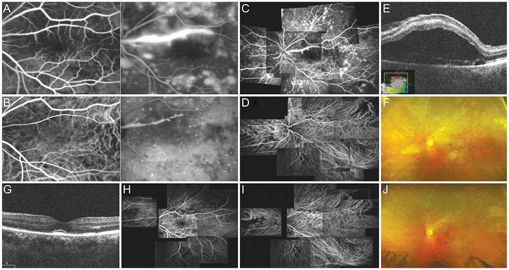
Fig. 1 (A) Fluorescein angiography (FA) showed delayed filling of the choroidal watershed zone and cilioretinal artery (CA) simultaneously with normal filling of arterial branches from the central retinal artery. (B) Indocyanine green angiography (ICGA) also showed multiple patchy hypofluorescence signals involving the posterior pole. There was no apparent arteriovenous transit time delay (10 seconds). In the late phase, perivascular leakage along the CA is evident and multiple blot hyperfluorescence on FA and pinpoint hyperfluorescence on ICGA are noted. (C) Panoramic FA showing multiple patchy hyperfluorescence signals with a wedge-shaped pattern across the fundus. Dilatation and staining of the CA is also present. (D) Panoramic ICGA showing multiple hypofluorescence signals with multiple patchy choroidal filling defects. (E) Optical coherence tomography showing diffuse serous retinal detachment with pigment epithelial detachment (central retinal thickness [CRT], 834 µm). (F) Optomap showing multiple patchy whitenings of the outer retina and yellowish pigmentary changes at the level of the retinal pigment epithelium (RPE) across the fundus. (G) Three days after the intravitreous bevacizumab injection, OCT shows markedly improved serous retinal detachment (SRD; CRT, 269 µm). Ten days after the injection, (H) panoramic FA shows improvement of leakage, but geographic areas of hypofluorescence persist. (I) Panoramic ICGA shows no signs of any significant leakage from choroidal vessels, but multiple patchy choroidal filling defects remain. (J) Optomap shows improvement of SRD and RPE ischemic changes.
Reference
-
1. Hayreh SS. Segmental nature of the choroidal vasculature. Br J Ophthalmol. 1975; 59:631–648.2. Loeffler KU, Hayreh SS, Tso MO. The effects of simultaneous occlusion of the posterior ciliary artery and vortex veins: a histopathologic study. Arch Ophthalmol. 1994; 112:674–682.3. Vinores SA, Youssri AI, Luna JD, et al. Upregulation of vascular endothelial growth factor in ischemic and non-ischemic human and experimental retinal disease. Histol Histopathol. 1997; 12:99–109.4. Kaur C, Sivakumar V, Yong Z, et al. Blood-retinal barrier disruption and ultrastructural changes in the hypoxic retina in adult rats: the beneficial effect of melatonin administration. J Pathol. 2007; 212:429–439.5. Kim M, Kwon HJ, Lee SC. Spontaneous resolution of posterior ciliary artery occlusion. Graefes Arch Clin Exp Ophthalmol. 2013; 251:1005–1006.
- Full Text Links
-
- Actions
-
Cited
- CITED
-
- Close
- Share
-
- Similar articles
-
- Serous Retinal Detachment Following Combined Photodynamic Therapy and Intravitreal Bevacizumab Injection
- A Case of Intravitreal Bevacizumab Injection for the Treatment of Choroidal Neovascularization in Morning Glory Syndrome
- Takayasu's Arteritis Associated with Serous Retinal Detachment
- The Short-term Effect of Intravitreal Bevacizumab for Treatment of Central Serous Chorioretinopathy
- Laser Photocoaculation Treatment in a Case of Circumscribged Choroidal hmangioma Associated with Serous Retinal Detachment
