Korean J Urol.
2015 Oct;56(10):717-721. 10.4111/kju.2015.56.10.717.
Distribution of ureteral stones and factors affecting their location and expulsion in patients with renal colic
- Affiliations
-
- 1Department of Urology, Konyang University College of Medicine, Daejeon, Korea. hjkim@kyuh.ac.kr
- KMID: 2344115
- DOI: http://doi.org/10.4111/kju.2015.56.10.717
Abstract
- PURPOSE
To evaluate the distribution of ureteral stones and to determine their characteristics and expulsion rate based on their location.
MATERIALS AND METHODS
We retrospectively reviewed computed tomography (CT) findings of 246 patients who visited our Emergency Department (ED) for renal colic caused by unilateral ureteral stones between January 2013 and April 2014. Histograms were constructed to plot the distribution of stones based on initial CT findings. Data from 144 of the 246 patients who underwent medical expulsive therapy (MET) for 2 weeks were analyzed to evaluate the factors responsible for the stone distribution and expulsion.
RESULTS
The upper ureter and ureterovesical junction (UVJ) were 2 peak locations at which stones initially lodged. Stones lodged at the upper ureter and ureteropelvic junction (group A) had a larger longitudinal diameter (4.21 mm vs. 3.56 mm, p=0.004) compared to those lodged at the lower ureter and UVJ (group B). The expulsion rate was 75.6% and 94.9% in groups A and B, respectively. There was no significant difference in the time interval from initiation of renal colic to arrival at the ED between groups A and B (p=0.422). Stone diameter was a significant predictor of MET failure (odds ratio [OR], 1.795; p=0.005) but the initial stone location was not (OR, 0.299; p=0.082).
CONCLUSIONS
The upper ureter and UVJ are 2 peak sites at which stones lodge. For stone size 10 mm or less, initial stone lodge site is not a significant predictor of MET failure in patients who have no previous history of active stone treatment in the ureter.
MeSH Terms
-
Adult
Female
Humans
Kidney Pelvis/pathology
Male
Middle Aged
Renal Colic/drug therapy/*pathology/radiography
Retrospective Studies
Sulfonamides/therapeutic use
Tomography, X-Ray Computed
Treatment Failure
Ureter/pathology
Ureteral Calculi/drug therapy/*pathology/radiography
Urological Agents/therapeutic use
Sulfonamides
Urological Agents
Figure
-
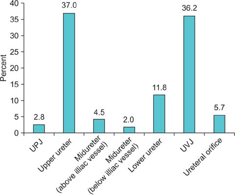
Fig. 1 The locations of ureteral stones in the anatomical portions of the ureter in 246 patients (total cohort). UPJ, ureteropelvic junction; UVJ, ureterovesical junction.
Reference
-
1. Stamatelou KK, Francis ME, Jones CA, Nyberg LM, Curhan GC. Time trends in reported prevalence of kidney stones in the United States: 1976-1994. Kidney Int. 2003; 63:1817–1823.2. Song HJ, Cho ST, Kim KK. Investigation of the location of the ureteral stone and diameter of the ureter in patients with renal colic. Korean J Urol. 2010; 51:198–201.3. Ketabchi AA, Mehrabi S. The effect of tamsulosin, an alpha-1 receptor antagonist as a medical expelling agent in success rate of ureteroscopic lithotripsy. Nephrourol Mon. 2013; 6:e12836.4. Chua ME, Gomez OR, Sapno LD, Lim SL, Morales ML Jr. Use of computed tomography scout film and Hounsfield unit of computed tomography scan in predicting the radio-opacity of urinary calculi in plain kidney, ureter and bladder radiographs. Urol Ann. 2014; 6:218–223.5. Ryu JA, Kim B, Jeon YH, Lee J, Lee JW, Jeon SS, et al. Unenhanced spiral CT in acute ureteral colic: a replacement for excretory urography? Korean J Radiol. 2001; 2:14–20.6. El-Barky E, Ali Y, Sahsah M, Terra AA, Kehinde EO. Site of impaction of ureteric calculi requiring surgical intervention. Urolithiasis. 2014; 42:67–73.7. Ordon M, Schuler TD, Ghiculete D, Pace KT, Honey RJ. Stones lodge at three sites of anatomic narrowing in the ureter: clinical fact or fiction? J Endourol. 2013; 27:270–276.8. Eisner BH, Reese A, Sheth S, Stoller ML. Ureteral stone location at emergency room presentation with colic. J Urol. 2009; 182:165–168.9. Bader MJ, Eisner B, Porpiglia F, Preminger GM, Tiselius HG. Contemporary management of ureteral stones. Eur Urol. 2012; 61:764–772.10. Jellison FC, Smith JC, Heldt JP, Spengler NM, Nicolay LI, Ruckle HC, et al. Effect of low dose radiation computerized tomography protocols on distal ureteral calculus detection. J Urol. 2009; 182:2762–2767.11. Park SH, Kim KD, Moon YT, Myung SC, Kim TH, Chang IH, et al. Pilot study of low-dose nonenhanced computed tomography with iterative reconstruction for diagnosis of urinary stones. Korean J Urol. 2014; 55:581–586.12. Eisner BH, Pedro R, Namasivayam S, Kambadakone A, Sahani DV, Dretler SP, et al. Differences in stone size and ureteral dilation between obstructing proximal and distal ureteral calculi. Urology. 2008; 72:517–520.13. Coll DM, Varanelli MJ, Smith RC. Relationship of spontaneous passage of ureteral calculi to stone size and location as revealed by unenhanced helical CT. AJR Am J Roentgenol. 2002; 178:101–103.14. Hubner WA, Irby P, Stoller ML. Natural history and current concepts for the treatment of small ureteral calculi. Eur Urol. 1993; 24:172–176.
- Full Text Links
-
- Actions
-
Cited
- CITED
-
- Close
- Share
-
- Similar articles
-
- The Effect of Tamsulosin on Expulsion of Ureteral Stones after Extracorporeal Shock Wave Lithotripsy
- Investigation of the Location of the Ureteral Stone and Diameter of the Ureter in Patients with Renal Colic
- Extracorporeal shock wave lithotripsy of ureteral stones : Investigation of the factors influencing upon stone fragmentation
- Factors affecting the urologist’s decision to administer ureteral stone therapy: a retrospective cohort study
- Management of Ureteral Stones
