J Korean Ophthalmol Soc.
2014 Jun;55(6):887-890.
Quantitative Measurement of the Sella Turcica in Pseudotumor Cerebri
- Affiliations
-
- 1Department of Ophthalmology, Dankook University Medical College, Cheonan, Korea. kseeye@hanmail.net
Abstract
- PURPOSE
In this study we evaluated the hypothesis that sella turcica enlarged in size due to increased intracranial hypertension by measuring the sella turcica area using magnetic resonance imaging (MRI) in patients with increased intracranial hypertension and compared to normal controls.
METHODS
Brain magnetic resonance (MR) midsagittal images of patients diagnosed with pseudotumor cerebri from 2005 to 2012 at Dankook University Hospital and 10 normal controls who had no overt signs or symptoms of neurological disease and had normal gadolinium-enhanced MR examination of brain were compared. The area of the sella turcica was measured by the double-blind method using Dicomworks v 1.3.5b (Philippe Puech and Loic Boussel, Freeware, France). Statistical analysis was conducted using GraphPad Prism (GraphPad Software, Inc., USA) and Mann-Whitney U-test.
RESULTS
The sella turcica areas of 2 pseudotumor cerebri patients were 93 mm2 and 123 mm2 and were significantly larger than in the controls (p = 0.03).
CONCLUSIONS
Empty sella which commonly occurs in pseudotumor cerebri can be caused by pituitary gland atrophy but, conversely, can result from the enlargement of the bony sella in response to an abnormal cerebrospinal fluid pressure gradient.
MeSH Terms
Figure
-
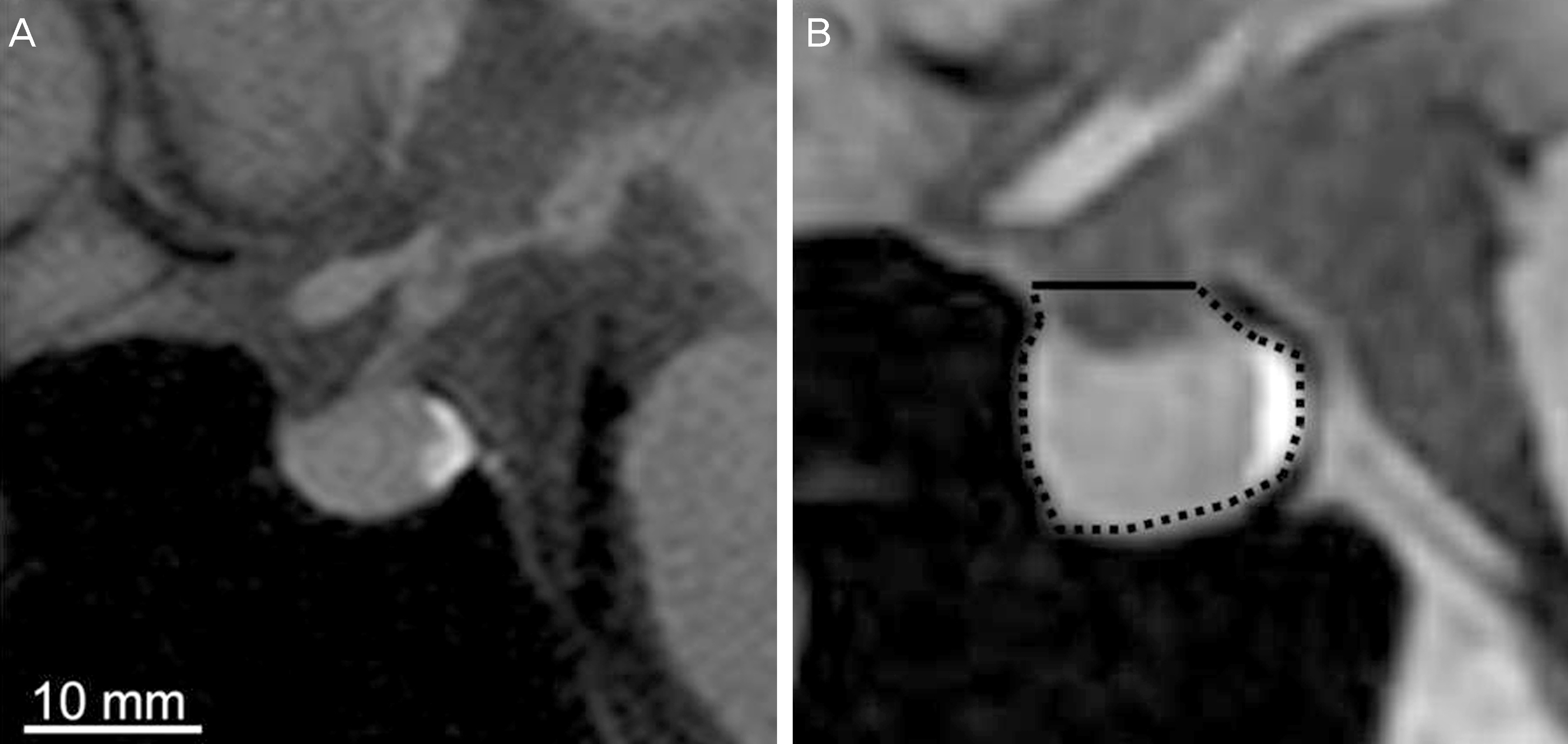
Figure 1. (A) T1-weighted mid-sagittal image of the sella turcica and pituitary gland from a normal subject. (B) Drawing showing the dimensions that were measured to determine the opening (solid line) and contour (dotted line) of the sella turcica. In this example, the sella area = 93 mm.
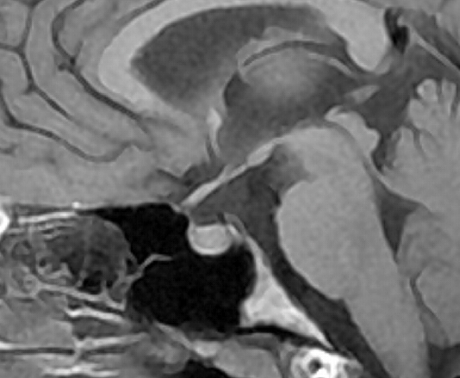
Figure 2. Tl-weighted mid-sagittal image of 17-year-old wom-an patient showed partially empty sella (turcica area: 93 mm2).
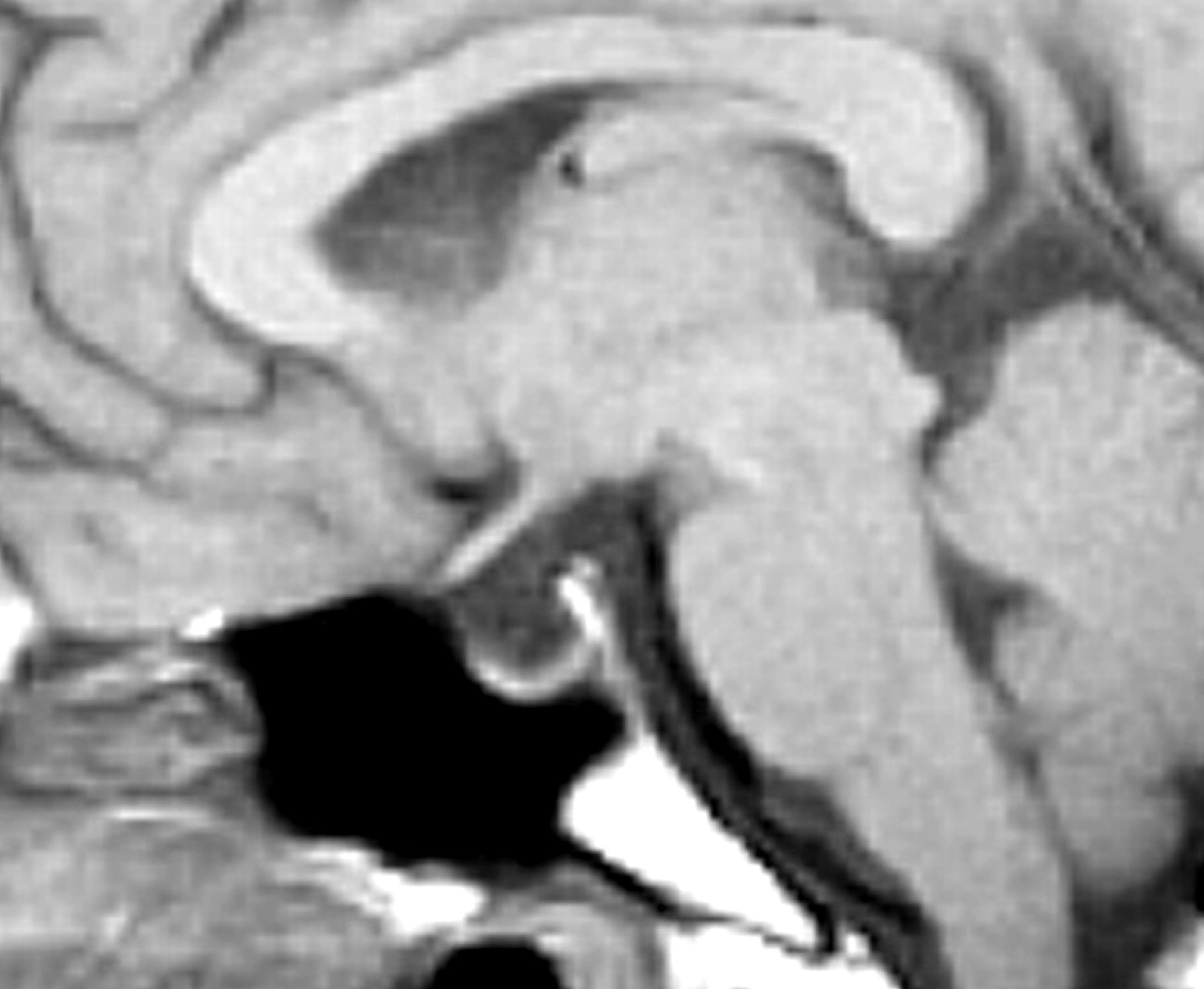
Figure 3. Tl-weighted mid-sagittal image of 28-year-old wom-an patient showed prominent empty sella (turcica area: 123 mm2).

Figure 4. The size of pseudotumor cerebri patients’ sella turcia was each 123 mm2, 93 mm2 and the median value was 108 mm2. The median value of the control group's sella turcia size was 67.5 mm2.
Reference
-
References
1. Brodsky MC, Vaphiades M. Magnetic resonance imaging in pseu-dotumor cerebri. Ophthalmology. 1998; 105:1686–93.
Article2. Park JW, Cha SH, Song GS, Choi CH. Pseudotumor cerebri asso-ciated with polycythemia vera. J Korean Neurosurg Soc. 2003; 34:162–4.3. Takanashi J, Suzuki H, Nagasawa K, et al. Empty sella in children as a key for diagnosis. Brain and Development. 2001; 23:422–3.
Article4. Messina D, Bono F, Fera F, et al. Empty sella and bilateral trans-verse sinus stenosis predict raised intracranial pressure in the ab-sence of papilloedema. J Neurol. 2006; 253:674–6.
Article5. Wessel K, Thron A, Linden D, et al. Pseudotumor cerebri: clinical and neuroradiological findings. Eur Arch Psychiatry Neurol Sci. 1987; 237:54–60.6. Kaufman B. The “empty” sella turcica–a manifestation of the intrasellar subarachnoid space. Radiology. 1968; 90:931–41.
Article7. Friedman DI, Jacobson DM. Diagnostic criteria for idiopathic in-tracranial hypertension. Neurology. 2002; 59:1492–5.
Article8. Yuh WT, Zhu M, Taoka T, et al. MR imaging of pituitary morphol-ogy in idiopathic intracranial hypertension. J Magn Reson Imaging. 2000; 12:808–13.
Article9. Davis PC, Hoffman JC Jr, Spencer T, et al. MR imaging of pituitary adenoma: CT, clinical, and surgical correlation. AJR Am J Roentgenol. 1987; 148:797–802.
Article10. Weisberg LA, Numuguchi Y. Neuroimaging in neuroendocrine diseases. Neurol Clin. 1986; 4:783–800.
Article11. Hwang TN, Rofagha S, McDermott MW, et al. Sunken eyes, sag-ging brain syndrome: bilateral enophthalmos from chronic intra-cranial hypotension. Ophthalmology. 2011; 118:2286–95.
Article12. Thurtell MJ, Bruce BB, Newman NJ, Biousse V. An update on idi-opathic intracranial hypertension. Rev Neurol Dis. 2010; 7:e56–68.
- Full Text Links
-
- Actions
-
Cited
- CITED
-
- Close
- Share
-
- Similar articles
-
- The evaluation of sella turcica on the shape and volume in class III patients
- Assessment of the Relationship between Sella Turcica Morphology and Delayed Dental Age
- A Case of Pseudotumor Cerebri Associated with Primary Antiphospholipid Syndrome
- Pseudotumor Cerebri Associated with Polycythemia Vera
- The Ventriculoperitoneal Shunting in Pseudotumor Cerebri: Report of 2 Cases
