J Korean Ophthalmol Soc.
2013 Dec;54(12):1844-1855.
Comparison of Diagnostic Power Among OCT Parameters According to Peripapillary Atrophy in High Myopic Glaucoma
- Affiliations
-
- 1Department of Ophthalmology, Chungnam National University College of Medicine, Daejeon, Korea. kcs61@cnu.ac.kr
- 2Research Institute for Medical Science, Chungnam National University, Daejeon, Korea.
Abstract
- PURPOSE
To evaluate diagnostic power to detect glaucoma in high myopic eyes with peripapillary atrophy among optical coherence tomography (OCT) parameters.
METHODS
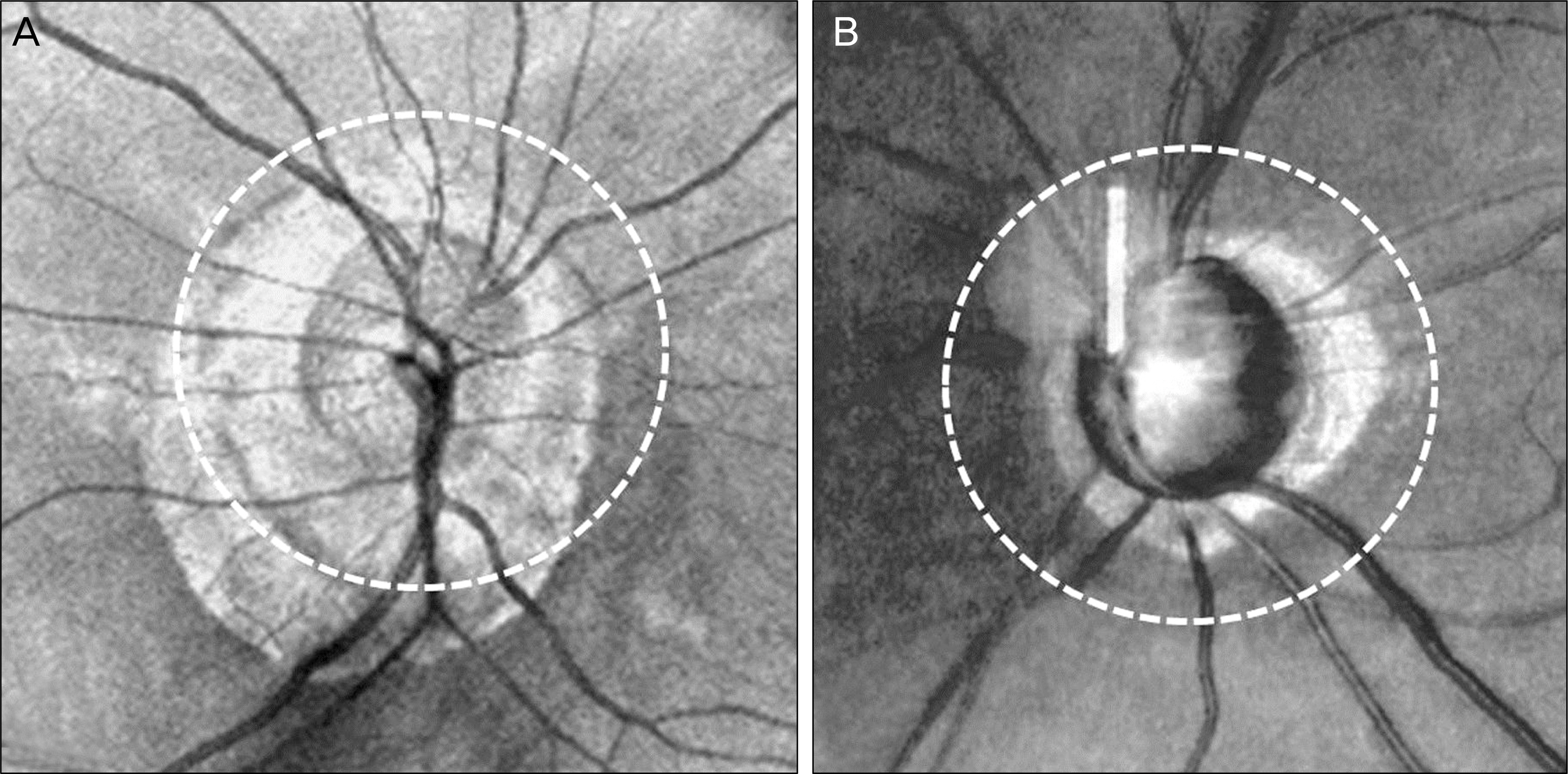
Fifty eyes of 31 glaucoma patients with myopia of -6.00 diopters or less and a peripapillary atrophy (PPA) were classified into a group with a PPA located beyond the circumpapillary OCT scan circle (group A) and a group with a PPA confined within the scan circle (group B). Circumpapillary retinal nerve fiber layer (cpRNFL), total macula (TM), and ganglion cell-inner plexiform layer (GCIPL) thickness were measured in each group and the diagnostic power of each measurement was compared by area under the receiver operating characteristic curve (AUC).
RESULTS
There were no significant differences in the age, gender, intraocular pressure, optic disc size, and mean deviation between the 2 groups. The spherical equivalent of group A was significantly larger than group B (mean -11.9 vs. -7.3 diopters, p = 0.002). In group A, the AUC of average GCIPL thickness was significantly higher than average cpRNFL and average TM thickness (p < 0.05). Additionally, when comparing parameters that showed the highest AUC value in each method, the AUC of GCIPL thickness was significantly higher than cpRNFL thickness (p = 0.046). In subgroup analysis of spherical equivalent matching between the 2 groups (subgroup A and B), the highest AUC value of GCIPL thickness was significantly higher than cpRNFL and TM thickness in subgroup A (p < 0.05). In group B and subgroup B, there was no statistical significance among AUC values of the 3 different methods (p > 0.05).
CONCLUSIONS
Assessment of GCIPL parameters is a useful technique for glaucoma diagnosis in patients with high myopia and PPA extending beyond circumpapillary OCT scan circle.
MeSH Terms
Figure
Reference
-
References
1. Tomlinson A, Phillips CI. Ratio of optic cup to optic disc: in rela-tion to axial length of eyeball and refraction. Br J Ophthamol. 1969; 53:765–8.
Article2. Chihara E, Sawada A. Atypical nerve fiber layer defects in high myopes with high-tension glaucoma. Arch Ophthalmol. 1990; 108:228–32.
Article3. Cahane M, Bartov E. Axial length and scleral thickness effect on susceptibility to glaucomatous damage: a theoretical model implementing Laplace's law. Ophthalmic Res. 1992; 24:280–4.
Article4. Avetisov ES, Savitskaya NF. Some features of ocular micro-circulation in myopia. Ann Ophthalmol. 1977; 9:1261–4.5. Shih YF, Horng IH, Yang CH. . Ocular pulse amplitude in myopia. J Ocul Pharmacol. 1991; 7:83–7.
Article6. To'mey KF, Faris BM, Jalkh AE, Nasr AM. Ocular pulse in high myopia: A study of 40 eyes. Ann Ophthalmol. 1981; 13:569–71.7. Ozdek SC, Onol M, Gürelik G, Hasanreisoglu B. Scanning laser polarimetry in normal subjects and patients with myopia. Br J Ophthalmol. 2000; 84:264–7.
Article8. Tay E, Seah SK, Chan SP. . Optic disk ovality as an index of tilt and its relationship to myopia and perimetry. Am J Ophthalmol. 2005; 139:247–52.
Article9. Chen TC, Cense B, Pierce MC. . Spectral domain optical co-herence tomography: ultra-high speed, ultra-high resolution oph-thalmic imaging. Arch Ophthalmol. 2005; 123:1715–20.10. Melo GB, Libera RD, Barbosa AS. . Comparison of optic disk and retinal nerve fiber layer thickness in nonglaucomatous and glaucomatous patients with high myopia. Am J Ophthalmol. 2006; 142:858–60.
Article11. Leung CK, Cheng AC, Chong KK. . Optic disc measurements in myopia with optical coherence tomography and confocal scan-ning laser ophthalmoscopy. Invest Ophthalmol Vis Sci. 2007; 48:3178–83.
Article12. Jonas JB, Nguyen XN, Gusek GC, Naumann GO. Parapapillary chorioretinal atrophy in normal and glaucoma eyes. I: Morphometric data. Invest Ophthalmol Vis Sci. 1989; 30:908–18.13. Jonas JB, Naumann GO. Parapapillary chorioretinal atrophy in normal and glaucoma eyes. II: Correlations. Invest Ophthalmol Vis Sci. 1989; 30:919–26.14. Jonas JB, Fernández MC, Naumann GO. Glaucomatous para-papillary atrophy. Occurrence and correlations. Arch Ophthalmol. 1992; 110:214–22.15. Park KH, Tomita G, Liou SY, Kitazawa Y. Correlation between peripapillary atrophy and optic nerve damage in normal-tension glaucoma. Ophthalmology. 1996; 103:1899–906.
Article16. Quigley HA, Dunkelberger GR, Green WR. Retinal ganglion cell atrophy correlated with automated perimetry in human eyes with glaucoma. Am J Ophthalmol. 1989; 107:453–64.
Article17. Zeimer R, Asrani S, Zou S. . Quantitative detection of glau-comatous damage at the posterior pole by retinal thickness mapping. A pilot study. Ophthalmology. 1998; 105:224–31.18. Greenfield DS, Bagga H, Knighton RW. Macular thickness changes in glaucomatous optic neuropathy detected using optical coherence tomography. Arch Ophthalmol. 2003; 121:41–6.
Article19. Leung CK, Chan WM, Yung WH. . Comparison of macular and peripapillary measurements for the detection of glaucoma: an optical coherence tomography study. Ophthalmology. 2005; 112:391–400.20. Tan O, Li G, Lu AT. . Advanced Imaging for Glaucoma Study Group. Mapping of macular substructures with optical coherence tomography for glaucoma diagnosis. Ophthalmology. 2008; 115:949–56.21. Kim NR, Lee ES, Seong GJ. . Comparing the ganglion cell complex and retinal nerve fiber layer measurements by Fourier do-main OCT to detect glaucoma in high myopia. Br J Ophthalmol. 2011; 95:1115–21.22. Shoji T, Nagaoka Y, Sato H, Chihara E. Impact of high myopia on the performance of SD-OCT parameters to detect glaucoma. Graefes Arch Clin Exp Ophthalmol. 2012; 250:1843–9.
Article23. Nonaka A, Hangai M, Akagi T. . Biometric features of peri-papillary atrophy beta in eyes with high myopia. Invest Ophthalmol Vis Sci. 2011; 52:6706–13.
Article24. O'Donnell C, Hartwig A, Radhakrishnan H. Correlations between refractive error and biometric parameters in human eyes using the LenStar 900. Cont Lens Anterior Eye. 2011; 34:26–31.25. Hoh ST, Lim MC, Seah SK. . Peripapillary retinal nerve fiber layer thickness variations with myopia. Ophthalmology. 2006; 113:773–7.
Article26. Hirasawa H, Tomidokoro A, Araie M. . Peripapillary retinal nerve fiber layer thickness determined by spectral-domain optical coherence tomography in ophthalmologically normal eyes. Arch Ophthalmol. 2010; 128:1420–6.
Article27. Leung CK, Mohamed S, Leung KS. . Retinal nerve fiber layer measurements in myopia: An optical coherence tomography study. Invest Ophthalmol Vis Sci. 2006; 47:5171–6.
Article28. Rauscher FM, Sekhon N, Feuer WJ, Budenz DL. Myopia affects retinal nerve fiber layer measurements as determined by optical co-herence tomography. J Glaucoma. 2009; 18:501–5.
Article29. Na JH, Moon BG, Sung KR. . Characterization of peripapillary atrophy using spectral domain optical coherence tomography. Korean J Ophthalmol. 2010; 24:353–9.
Article30. Manjunath V, Shah H, Fujimoto JG, Duker JS. Analysis of peri-papillary atrophy using spectral domain optical coherence tomography. Ophthalmology. 2011; 118:531–6.
Article31. Kang SH, Hong SW, Im SK. . Effect of myopia on the thickness of the retinal nerve fiber layer measured by Cirrus HD optical coher-ence tomography. Invest Ophthalmol Vis Sci. 2010; 51:4075–83.
Article32. Lam DS, Leung KS, Mohamed S. . Regional variations in the relationship between macular thickness measurements and myopia. Invest Ophthalmol Vis Sci. 2007; 48:376–82.
Article33. Choi SW, Lee SJ. Thickness changes in the fovea and peripapillary retinal nerve fiber layer depend on the degree of myopia. Korean J Ophthalmol. 2006; 20:215–9.
Article34. Kim SH, Park JY, Park TK, Ohn YH. Use of spectral-domain opti-cal coherence tomography to analyze macular thickness according to refractive error. J Korean Ophthalmol Soc. 2011; 52:1286–95.
Article
- Full Text Links
- Actions
-
Cited
- CITED
-
- Close
- Share
- Similar articles
-
- Influence of Peripapillary Atrophy on the Progress of Diabetic Retinopathy
- Peripapillary Atrophy in Asymmetric Glaucoma
- Comparison of Peripapillary Atrophy between Normal Tension Glaucoma and Glaucoma-like Disc
- Additive Role of Optical Coherence Tomography Angiography Vessel Density Measurements in Glaucoma Diagnoses
- Diagnosis of Glaucoma in the Cases of the Opposed Results of GDx and OCT


