A Case of Serous Macular Detachment Associated with Tractional Fibrous Tissue in an Optic Pit Patient
- Affiliations
-
- 1Department of Ophthalmology, Konyang University, Kim's Eye Hospital, Myung-Gok Eye Research Institute, Seoul, Korea. eyecure@kimeye.com
Abstract
- PURPOSE
To report a case of serous macular detachment associated with tractional fibrous tissue in an optic pit patient successfully treated by vitrectomy without laser photocoagulation.
CASE SUMMARY
A 15-year-old female visited our hospital for visual disturbance of her left eye. The patient's best-corrected visual acuity was 0.5. After ophthalmic examinations, the patient was diagnosed with an optic pit associated serous macular detachment. Pars plana vitrectomy with complete posterior vitreous detachment was performed. A thick fibrous tissue within the optic disc cupping was found. During removal of the fibrous tissue, the tissue was observed to be attached to a tiny hole above the optic pit. The thick fibrous tissue was tugging at the margin of the hole and was removed using intraocular forceps to relieve the traction. Fluid-gas exchange was then performed and the operation was completed without laser photocoagulation around the optic disc. The fovea was reattached completely in 12 months and visual acuity was improved to 0.8.
MeSH Terms
Figure
-

Figure 1 Preoperative fundus photographs shows optic disc pit in temporal area and about 3D size serous retinal detachment.
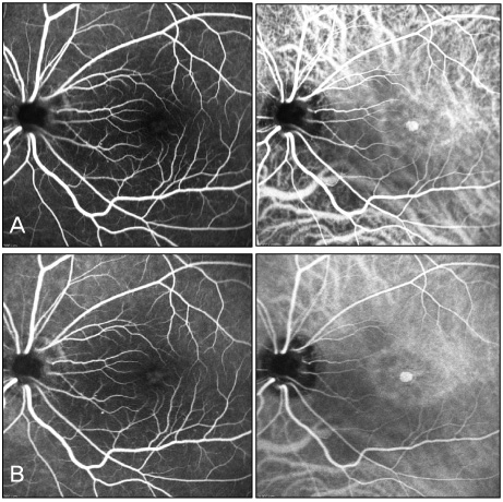
Figure 2 Preoperative FAG and HRA show hyperfluorescence in macular area in early phase (A) and pooling of fluorescein in macular area in late phase (B).

Figure 3 Preoperative OCT (A) shows retinoschisis and subretinal fluid at the macular. The retinoschisis extended to the optic disc pit. Preoperative macular thickness was 772 µm on topography (B).

Figure 4 Thick fibrous tissue (arrow) existed on the cup of the optic nerve head (A). We found a tiny hole (arrow) above the optic pit during removal of the thick fibrous tissue (B).

Figure 5 Postoperative OCT shows decreased subretinal fluind and retinoschisis. (A) POD # 3 month, (B) POD # 6 month, (C) POD # 9 month, (D) POD # 12 month.
Reference
-
1. Brown GC, Shields JA, Goldberg RE. Congenital pits of the optic nerve head. II. Clinical studies in humans. Ophthalmology. 1980. 87:51–65.2. Lincoff H, Lopez R, Kreissig I, et al. Retinoschisis associated with optic nerve pits. Arch Ophthalmol. 1988. 106:61–67.3. Lincoff H, Kreissig I. Optical coherence tomography of pneumatic displacement of optic disc pit maculopathy. Br J Ophthalmol. 1998. 82:367–372.4. Rutledge BK, Puliafito CA, Duker JS, et al. Optical coherence tomography of macular lesions associated with optic nerve head pits. Ophthalmology. 1996. 103:1047–1053.5. Brockhurst RJ. Optic pits and posterior retinal detachment. Trans Am Ophthalmol Soc. 1975. 73:264–291.6. Schneider M, Geitzenauer W, Ahlers C, et al. Three-dimensional imaging of an optic disk pit using high resolution optical coherence tomography. Eur J Ophthalmol. 2009. 19:321–323.7. Savell J, Cook JR. Optic nerve colobomas of autosomal-dominant heredity. Arch Ophthalmol. 1976. 94:395–400.8. Kalina RE, Conrad WC. Letter: Intrathecal fluorescein for serous macular detachment. Arch Ophthalmol. 1976. 94:1421.9. Georgalas I, Kouri A, Ladas I, Gotzaridis E. Optic disc pit maculopathy treated with vitrectomy, internal limiting membrane peeling, and air in a 5-year-old boy. Can J Ophthalmol. 2010. 45:189–191.10. Hirakata A, Okada AA, Hida T. Long-term results of vitrectomy without laser treatment for macular detachment associated with an optic disc pit. Ophthalmology. 2005. 112:1430–1435.11. Brown GC, Shields JA, Patty BE, Goldberg RE. Congenital pits of the optic nerve head. I. Experimental studies in collie dogs. Arch Ophthalmol. 1979. 97:1341–1344.12. Maia OO Jr, Soriano DS, Takahashi WY, Suzuki H. Surgical treatment of macular detachment secondary to congenital pit of the optic disc: case report. Arq Bras Oftalmol. 2008. 71:874–877.13. Ryu JW, Ra H, Lee WK. A case of surgically treated serous macular detachment associated with optic disc pit. J Korean Ophthalmol Soc. 2010. 51:155–158.14. Georgalas I, Petrou P, Koutsandrea C, et al. Optic disc pit maculopathy treated with vitrectomy, internal limiting membrane peeling, and gas tamponade: a report of two cases. Eur J Ophthalmol. 2009. 19:324–326.15. Dai S, Polkinghorne P. Peeling the internal limiting membrane in serous macular detachment associated with congenital optic disc pit. Clin Experiment Ophthalmol. 2003. 31:272–275.16. Inoue M, Shinoda K, Ishida S. Vitrectomy combined with glial tissue removal at the optic pit in a patient with optic disc pit maculopathy: a case report. J Med Case Reports. 2008. 2:103.17. Akiba J, Kakehashi A, Hikichi T, Trempe CL. Vitreous findings in cases of optic nerve pits and serous macular detachment. Am J Ophthalmol. 1993. 116:38–41.
- Full Text Links
-
- Actions
-
Cited
- CITED
-
- Close
- Share
-
- Similar articles
-
- A Case of Optic Disc Pit
- A Case of Serous Macular Detachment Preceding Macular Retinoschisis in an Optic Pit
- A Case of Surgically Treated Serous Macular Detachment Associated With Optic Disc Pit
- A Case of Optic Pit Maculopathy Treated with Fovea-sparing and an Inverted Flap Technique
- Case of Macular Hole after Surgery in Macular Detachment with Optic Disc Pit in a Child
