J Korean Ophthalmol Soc.
2012 Mar;53(3):365-371.
Clinical Outcomes of Advanced Surface Ablation with Smoothing in High Myopia
- Affiliations
-
- 1Lee Eye Clinic, Busan, Korea. eyebong1@hanmail.net
- 2Department of Ophthalmology, Kosin University College of Medicine, Busan, Korea.
Abstract
- PURPOSE
To investigate the results of Advanced Surface Ablation (ASA) coupled with "smoothing" to smooth the ablation surface after covering masking fluid.
METHODS
ASA was performed in 61 eyes with ablation depth of more than 75 microm. The mean refractive error was -5.88 +/- 1.27 D and mean ablation depth was 102.93 +/- 12.06 microm. Smoothing was performed in all patients (mean depth 16.79 +/- 2.43 microm, mean diameter 8.77 +/- 0.16 mm). Customized Aspheric Transition zone (CATz) was used in the laser algorithm.
RESULTS
The mean refractive error was -0.29 +/- 0.41 D at postoperative 6 months and 97% of patients had an UCVA of 1.0 or better at postoperative 6 months. There was no statistically significant difference in magnitude of high-order aberrations at postoperative 6 months. The magnitude of total spherical aberrations increase was 0.084 microm at postoperative 6 months (p<0.05, paired t-test). The cornea was maintained clear in the majority of eyes.
CONCLUSIONS
Excellent results were obtained by ASA coupled with "smoothing" in high myopia patients with an ablation depth greater than 75 microm.
Keyword
Figure
-
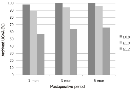
Figure 1 Distribution of uncorrected visual acuity after advanced surface ablation with smoothing over time.
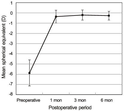
Figure 2 Mean spherical equivalent following advanced surface ablation with smoothing at different follow-up periods.
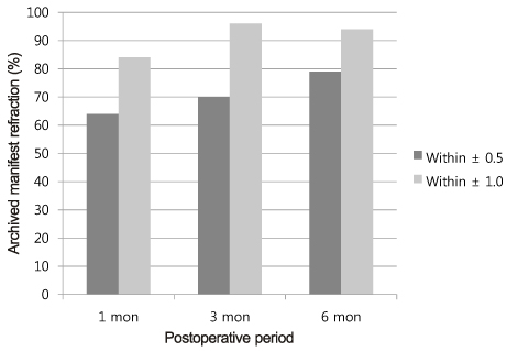
Figure 3 Distribution of eyes in a given range of emmetropia after advanced surface ablation with smoothing.
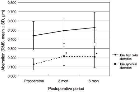
Figure 4 Changes in total high order aberration and total spherical aberration during postoperative 6 months. *p < 0.05 (paired t-test) compared with preoperative value.
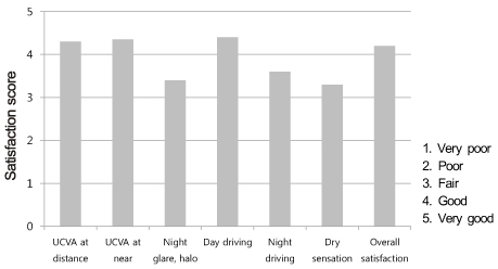
Figure 5 Patient's satisfaction after advanced surface ablation with smoothing.
Reference
-
1. Rao SK, Srinivasan B, Sitalakshmi G, Padmanabhan P. Photorefractive keratectomy versus laser in situ keratomileusis to prevent keratectasia after corneal ablation. J Cataract Refract Surg. 2004. 30:2623–2628.2. Lee SB, Chung MS. Advanced Surface Ablation-Photorefractive Keratectomy (ASA-PRK): Safety and clinical outcome for the correction of mild to moderate myopia with a thin cornea. J Korean Ophthalmol Soc. 2006. 47:1274–1286.3. Randleman JB, Loft ES, Banning CS, et al. Outcomes of wave-front-optimized surface ablation. Ophthalmology. 2007. 114:983–988.4. Ghadhfan F, Al-Rajhi A, Wagoner MD. Laser in situ keratomileusis versus surface ablation: visual outcomes and complications. J Cataract Refract Surg. 2007. 33:2041–2048.5. Alió JL, Muftuoglu O, Ortiz D, et al. Ten-year follow-up of photorefractive keratectomy for myopia of more than -6 diopters. Am J Ophthalmol. 2008. 145:37–45.6. Lee JK, Choi WS, Choi YI. Excimer laser photorefractive keratectomy for high myopia. J Korean Ophthalmol Soc. 1994. 35:927–934.7. Carson CA, Taylor HR. Excimer laser treatment for high and extreme myopia. The Melbourne Excimer Laser and Research Group. Arch Ophthalmol. 1995. 113:431–436.8. Jeong JW, Hahn YH. Complications after excimer laser photorefractive keratectomy in high myopia. J Korean Ophthalmol Soc. 1997. 38:1128–1138.9. Wachtlin J, Langenbeck K, Schründer S, et al. Immunohistology of corneal wound healing after photorefractive keratectomy and laser in situ keratomileusis. wachtlin@ukbf.fu-berlin.de. J Refract Surg. 1999. 15:451–458.10. Lin N, Yee SB, Mitra S, et al. Prediction of corneal haze using an ablation depth/corneal thickness ratio after laser epithelial keratomileusis. J Refract Surg. 2004. 20:797–802.11. Virasch VV, Majmudar PA, Epstein RJ, et al. Reduced application time for prophylactic mitomycin C in photorefractive keratectomy. Ophthalmology. 2010. 117:885–889.12. Vinciguerra P, Azzolini M, Radice P, et al. A method for examining surface and interface irregularities after photorefractive keratectomy and laser in situ keratomileusis: predictor of optical and functional outcomes. J Refract Surg. 1998. 14:2 Suppl. S204–S206.13. Vinciguerra P, Azzolini M, Airahgi P, et al. Effect of decreasing surface and interface irregularities after photorefractive keratectomy and laser in situ keratomileusis on optical and functional outcomes. J Refract Surg. 1998. 14:2 Suppl. S199–S203.14. Maldonado MJ. Intraoperative MMC after excimer laser surgery for myopia. Ophthalmology. 2002. 109:826.15. Jain S, McCally RL, Connolly PJ, Azar DT. Mitomycin C reduces corneal light scattering after excimer keratectomy. Cornea. 2001. 20:45–49.16. Fantes FE, Hanna KD, Waring GO 3rd, et al. Wound healing after excimer laser keratomileusis (photorefractive keratectomy) in monkeys. Arch Ophthalmol. 1990. 108:665–675.17. Pallikaris IG, Papatzanaki ME, Stathi EZ, et al. Laser in situ keratomileusis. Lasers Surg Med. 1990. 10:463–468.18. Wang Z, Chen J, Yang B. Posterior corneal surface topographic changes after laser in situ keratomileusis are related to residual corneal bed thickness. Ophthalmology. 1999. 106:406–409.19. Joo CK, Kim TG. Corneal ectasia detected after laser in situ keratomileusis for correction of less than -12 diopters of myopia. J Cataract Refract Surg. 2000. 26:292–295.20. Pallikaris IG, Kymionis GD, Astyrakakis NI. Corneal ectasia induced by laser in situ keratomileusis. J Cataract Refract Surg. 2001. 27:1796–1802.21. Lee DH, Seo S, Jeong KW, et al. Early spatial changes in the posterior corneal surface after laser in situ keratomileusis. J Cataract Refract Surg. 2003. 29:778–784.22. Miyata K, Tokunaga T, Nakahara M, et al. Residual bed thickness and corneal forward shift after laser in situ keratomileusis. J Cataract Refract Surg. 2004. 30:1067–1072.23. Kim HJ, Cho SH, Kim JH, Joo CK. Risk factors and clinical evaluation for corneal ectasia after LASIK. J Korean Ophthalmol Soc. 2005. 46:589–596.24. Amoils SP, Deist MB, Gous P, Amoils PM. Iatrogenic keratectasia after laser in situ keratomileusis for less than -4.0 to -7.0 diopters of myopia. J Cataract Refract Surg. 2000. 26:967–977.25. Argento C, Cosentino MJ, Tytiun A, et al. Corneal ectasia after laser in situ keratomileusis. J Cataract Refract Surg. 2001. 27:1440–1448.26. Wang JC, Hufnagel TJ, Buxton DF. Bilateral keratectasia after unilateral laser in situ keratomileusis: a retrospective diagnosis of ectatic corneal disorder. J Cataract Refract Surg. 2003. 29:2015–2018.27. Lifshitz T, Levy J, Klemperer I, Levinger S. Late bilateral keratectasia after LASIK in a low myopic patient. J Refract Surg. 2005. 21:494–496.28. Carones F, Vigo L, Scandola E, Vacchini L. Evaluation of the prophylactic use of mitomycin-C to inhibit haze formation after photorefractive keratectomy. J Cataract Refract Surg. 2002. 28:2088–2095.29. Hashemi H, Taheri SM, Fotouhi A, Kheiltash A. Evaluation of the prophylactic use of mitomycin C to inhibit haze formation after photorefractive keratectomy in high myopia: a prospective clinical study. BMC Ophthalmol. 2004. 4:12.30. Camellin M. Laser epithelial keratomileusis with mitomycin C: Indications and limits. J Refract Surg. 2004. 20:5 Suppl. S693–S698.31. Gambato C, Ghirlando A, Moretto E, et al. Mitomycin C modulation of corneal wound healing after photorefractive keratectomy in highly myopic eyes. Ophthalmology. 2005. 112:208–218.32. Pfister RR. Permanent corneal edema resulting from the treatment of PTK corneal haze with mitomycin: a case report. Cornea. 2004. 23:744–747.33. Qazi MA, Johnson TW, Pepose JS. Development of late-onset subepithelial corneal haze after laser-assisted subepithelial keratectomy with prophylactic intraoperative mitomycin-C Case report and literature review. J Cataract Refract Surg. 2006. 32:1573–1578.34. Kim ES, Jin KH. Evaluation of the Prophylactic Use of Mitomycin to Inhibit Haze Formation after LASEK. J Korean Ophthalmol Soc. 2007. 48:623–629.35. Huang D, Tang M, Shekhar R. Mathematical model of corneal surface smoothing after laser refractive surgery. Am J Ophthalmol. 2003. 135:267–278.36. Balestrazzi E, De Molfetta V, Spadea L, et al. Histological, immunohistochemical, and ultrastructural findings in human corneas after photorefractive keratectomy. J Refract Surg. 1995. 11:181–187.37. Oshika T, Klyce SD, Applegate RA, et al. Comparison of corneal wavefront aberrations after photorefractive keratectomy and laser in situ keratomileusis. Am J Ophthalmol. 1999. 127:1–7.38. Kwon HL, Kim KI, Koo BS, Park HR. Short term clinical results of laser epithelial keratomileusis and epi-laser in situ keratomileusis for moderate and high myopia. J Korean Ophthalmol Soc. 2005. 46:1711–1717.39. Choi SK, Park HY, Kim YH, Chung SK. Comparison of laser epithelial keratomileusis versus epipolis-laser in situ keratomileusis for moderate to high myopia. J Korean Ophthalmol Soc. 2007. 48:1196–1201.40. Vinciguerra P, Camesasca FI, Randazzo A. One-year results of butterfly laser epithelial keratomileusis. J Refract Surg. 2003. 19:2 Suppl. S223–S226.41. Vinciguerra P, Torres I, Camesasca FI. Applications of confocal microscopy in refractive surgery. J Refract Surg. 2002. 18:3 Suppl. S378–S381.42. Vinciguerra P, Camesasca FI. Treatment of hyperopia: a new ablation profile to reduce corneal eccentricity. J Refract Surg. 2002. 18:3 Suppl. S315–S317.43. Serrao S, Lombardo M. One-year results of Photorefractive keratectomy with and without surface smoothing using the Technolas 217C laser. J Refract Surg. 2004. 20:444–449.44. Lee HK, Lee KS, Kim JK, et al. Epithelial healing and clinical outcomes in excimer laser photorefractive surgery following three epithelial removal techniques: mechanical, alcohol, and excimer laser. Am J Ophthalmol. 2005. 139:56–63.45. Vinciguerra P, Camesasca FI, Torres IM. Transition zone design and smoothing in custom laser-assisted subepithelial keratectomy. J Cataract Refract Surg. 2005. 31:39–47.46. Dougherty PJ, Waring G 3rd, Chayet A, et al. Topographically guided laser in situ keratomileusis for myopia using a customized aspherical treatment zone. J Cataract Refract Surg. 2008. 34:1862–1871.47. Vinciguerra P, Camesasca FI, Bains HS, et al. Photorefractive keratectomy for primary myopia using NIDEK topography-guided custumized aspheric transition zone. J Refract Surg. 2009. 25:1 Suppl. S89–S92.48. Kim JS, Lee SB. Effects of amount of myopic correction on long-term changes in higher-order wavefront aberrations in ASA-PRK. J Korean Ophthalmol Soc. 2010. 51:1184–1195.49. Lee SM, Lee MJ, Kim MK, et al. Comparison of changes in higher-order aberrations between conventional and wavefront-guided LASEK. J Korean Ophthalmol Soc. 2007. 48:1028–1035.
- Full Text Links
-
- Actions
-
Cited
- CITED
-
- Close
- Share
-
- Similar articles
-
- Clinical Outcomes of Advanced Surface Ablation with Smoothing
- Comparison of 10-year Clinical Results between Laser in situ Keratomileusis and Surface Ablation for Moderate to High Myopia
- Advanced Surface Ablation-Photorefractive Keratectomy (ASA-PRK): Safety and Clinical Outcome for the Correction of Mild to Moderate Myopia with a Thin Cornea
- Changes in Corneal Sensation, Tear Film Stability and Ocular Surface after Advanced Surface Ablation
- Simultaneous LASIK on both Stromal Surface and Flap Undersurface for High Myopia with Insufficient Corneal Bed
