Central Corneal Thickness Measured by Four Different Methods in Normal and Post-Femtosecond Laser-Assisted LASIK Eyes
- Affiliations
-
- 1Department of Ophthalmology, Ilsan Paik Hospital, Inje University College of Medicine, Goyang, Korea. jhk0924@hanmail.net
Abstract
- PURPOSE
To compare corneal pachymetry assessment using four measurement methods in normal and post-femtosecond laserassisted LASIK eyes.
METHODS
Central corneal thickness was measured sequentially using Orbscan II, Pentacam, Galilei and ultrasonic pachymetry in 30 normal, non-surgical eyes (Group I), 30 eyes one to six months after femtosecond laser-assisted LASIK (Group II), and 30 eyes six months or longer after femtosecond laser-assisted LASIK (Group III).
RESULTS
In Group I, corneal thickness measurements were similar for all four methods (P=0.202, one way ANOVA). In Groups II and III, corneal thickness measurements were significantly different (P=0.000, respectively, one way ANOVA). Compared to the Pentacam, Galilei and ultrasonic pachymetry, Orbscan significantly underestimated the corneal thicknesses in Groups II and III (P<0.005, respectively, one way ANOVA).
CONCLUSIONS
Central corneal thicknesses of normal eyes were similar for all four measurements, therefore corneal thickness measurements before refractive surgery using all four measurements is suitable. However measurements obtained with the Orbscan II were thinner than those obtained with the Pentacam, Galilei or ultrasonic pachymetry in post femtosecond laserassisted LASIK eyes. Further studies are needed to determine which instrument is more accurate in measuring central corneal thickness before and after refractive surgery.
MeSH Terms
Figure
-

Figure 1. Mean and 95% CI of CCT measurement by Orbscan, Pentacam, Galilei and US in Group I (CI=confidence interval; CCT=central.thickness; US=Ultrasonic pachymetry).

Figure 2. Mean and 95% CI of CCT Measurement by Orbscan, Pentacam, Galilei and US in Group II (CI= confidence interval; CCT=central. thickness; US= Ultrasonic pachymetry).
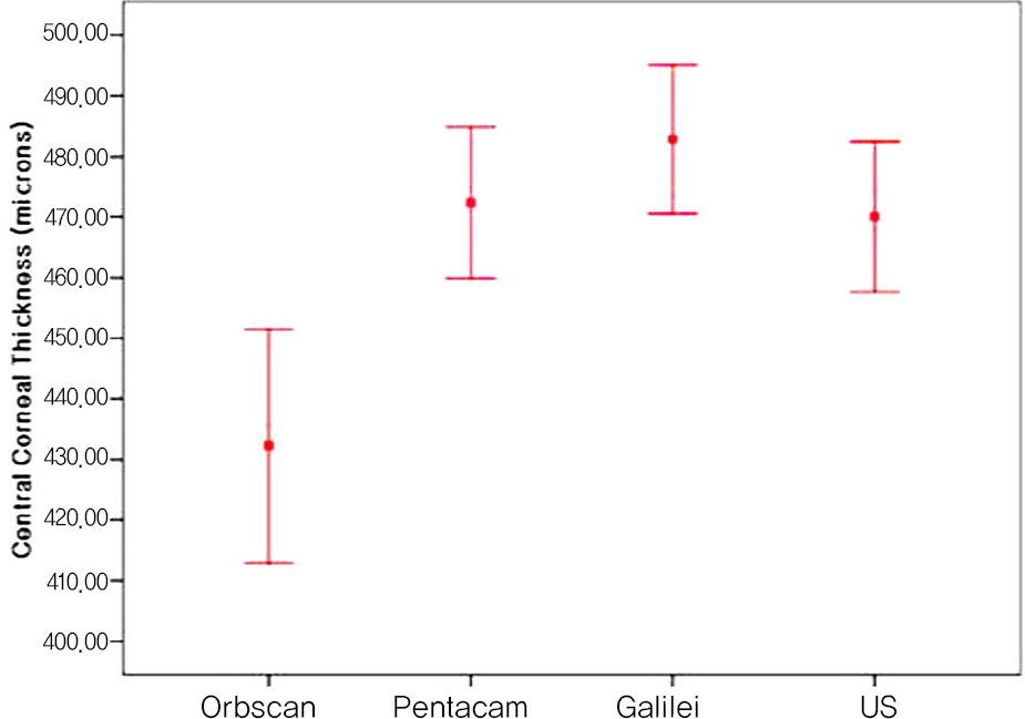
Figure 3. Mean and 95% CI of CCT Measurement by Orbscan, Pentacam, Galilei and US in Group III. (CI= confidence interval; CCT=central. thickness; US=Ultrasonic pachymetry).

Figure 4. Bland-Altman plots of Galilei, Pentacam and Orbscan CCT readings against US measurement in Group I. The middle line is the mean and the lines on the side represent the upper and lower 95% limits of agreement (LoA).

Figure 5. Bland-Altman plots of Galilei, Pentacam and Orbscan CCT readings against US measurement in Group II. The middle line is the mean and the lines on the side represent the upper and lower 95% limits of agreement (LoA).

Figure 6. Bland-Altman plots of Galilei, Pentacam and Orbscan CCT readings against US measurement in Group III. The middle line is the mean and the lines on the side represent the upper and lower 95% limits of agreement (LoA).
Reference
-
References
1. Binder PS. Ectasia after laser in situ keratomileusis. J Cataract Refract Surg. 2003; 29:2419–29.
Article2. Price FW Jr, Koller DL, Price MO. Central corneal pachymetry in patients undergoing laser in situ keratomileusis. Ophthalmology. 1999; 106:2216–20.3. Iskander NG, Peters NT, Penno EA, Gimbel HV. Postoperative complications in laser in situ keratomileusis. Curr Opin Ophthalmol. 2000; 11:273–9.
Article4. Miglior S, Albe E, Guareschi M, et al. Intraobserver and interobserver reproducibility in the evaluation of ultrasonic pachymetry measurements of central corneal thickness. Br J Ophthalmol. 2004; 88:174–7.
Article5. Solomon OD. Corneal indentation during ultrasonic pachometry. Cornea. 1999; 18:214–5.
Article6. Rufer F, Schroder A, Arvani MK, Erb C. Central and peripheral corneal pachymetry–standard evaluation with the Pentacam system. Klin Monatsbl Augenheilkd. 2005; 222:117–22.7. Menassa N, Kaufmann C, Goggin M, et al. Comparison and reproducibility of corneal thickness and curvature readings obtained by the Galilei and the Orbscan II analysis systems. J Cataract Refract Surg. 2008; 34:1742–47.
Article8. Kang PS, Kim JD, Yang YS. Comparison of corneal thickness measurements with the orbscan and ultrasonic pachymetry. J Korean Ophthalmol Soc. 2000; 41:1697–703.9. Kim SH, Cho JH, Song BJ. Accuracy of Orbscan Pachymetry Measurements and Ultrasonic Pachymetry before and after LASIK with Orbscan IIⓇ Topography. J Korean Ophthalmol Soc. 2002; 43:2513–8.10. Kwon M, Seoung Y, Hur D. Pachymetric measurements using orbscan after excimer refractive surgery. J Korean Ophthalmol Soc. 2004; 45:899–907.11. Shin YJ, Kim NH, Kim DH. Comparison of Pentacam with Orbscan. J Korean Ophthalmol Soc. 2007; 48:637–41.12. Ho T, Cheng AC, Rao SK, et al. Central corneal thickness measurements using Orbscan II, Visante, ultrasound, and Pentacam pachymetry after laser in situ keratomileusis for myopia. J Cataract Refract Surg. 2007; 33:1177–82.
Article13. Hashemi H, Mehravaran S. Central corneal thickness measurement with Pentacam, Orbscan II, and ultrasound devices before and after laser refractive surgery for myopia. J Cataract Refract Surg. 2007; 33:1701–7.
Article14. Kim SW, Byun YJ, Kim EK, Kim TI. Central corneal thickness measurements in unoperated eyes and eyes after PRK for myopia using Pentacam, Orbscan II, and ultrasonic pachymetry. J Refract Surg. 2007; 23:888–94.
Article15. Cheng AC, Rao SK, Tang E, Lam DS. Pachymetry assessment with Orbscan II in postoperative patients with myopic LASIK. J Refract Surg. 2006; 22:363–6.
Article16. Prisant O, Calderon N, Chastang P, et al. Reliability of pachymetric measurements using Orbscan after excimer refractive surgery. Ophthalmology. 2003; 110:511–5.
Article17. Yaylali V, Kaufman SC, Thompson HW. Corneal thickness measurements with the Orbscan Topography System and ultrasonic pachymetry. J Cataract Refract Surg. 1997; 23:1345–50.
Article18. Giessler S, Duncker GI. Orbscan pachymetry after LASIK is not reliable. J Refract Surg. 2001; 17:385–7.
Article19. Boscia F, La Tegola MG, Alessio G, Sborgia C. Accuracy of Orbscan optical pachymetry in corneas with haze. J Cataract Refract Surg. 2002; 28:253–8.
Article20. Fakhry MA, Artola A, Belda JI, et al. Comparison of corneal pachymetry using ultrasound and Orbscan II. J Cataract Refract Surg. 2002; 28:248–52.
Article21. von Jagow B, Kohnen T. Corneal architecture of femtosecond laser and microkeratome flaps imaged by anterior segment optical coherence tomography. J Cataract Refract Surg. 2009; 35:35–41.
Article
- Full Text Links
-
- Actions
-
Cited
- CITED
-
- Close
- Share
-
- Similar articles
-
- Reproducibility of IntraLASIK Flap Thickness Measured with Optical Coherence Tomography
- Influence of Central Corneal Thickness Change on Intraocular Pressure Measurement after LASIK and LASEK
- Comparison of Corneal Thickness Measurements Between Orbscan Topography System and Ultrasonic Pachymetry before and after LASIK
- Comparision of Corneal Endothelial Change in Femtosecond Laser and Microkeratome LASIK
- Factors Influencing Corneal Flap Thickness in Laser In Situ Keratomileusis with a Femtosecond Laser
