J Korean Ophthalmol Soc.
2008 Nov;49(11):1850-1856.
Two Cases of Giant Cell Angiofibroma in the Orbit
- Affiliations
-
- 1Department of Ophthalmology, Samsung Medical Center, Sungkyunkwan University School of Medicine, Seoul, Korea. ydkimoph@skku.edu
- 2Department of Ophthalmology, Dongguk University School of Medicine, Gyeongju, Korea.
- 3Department of Ophthalmology, Hallym University College of Medicine, Hallym University Sacred Heart Hospital, Seoul, Korea.
- 4Department of Pathology, Samsung Medical Center, Sungkyunkwan University School of Medicine, Seoul, Korea.
- 5Department of Radiology, Samsung Medical Center, Sungkyunkwan University School of Medicine, Seoul, Korea.
Abstract
- PURPOSE
To report two cases of giant cell angiofibroma in the orbit.
CASE SUMMARY
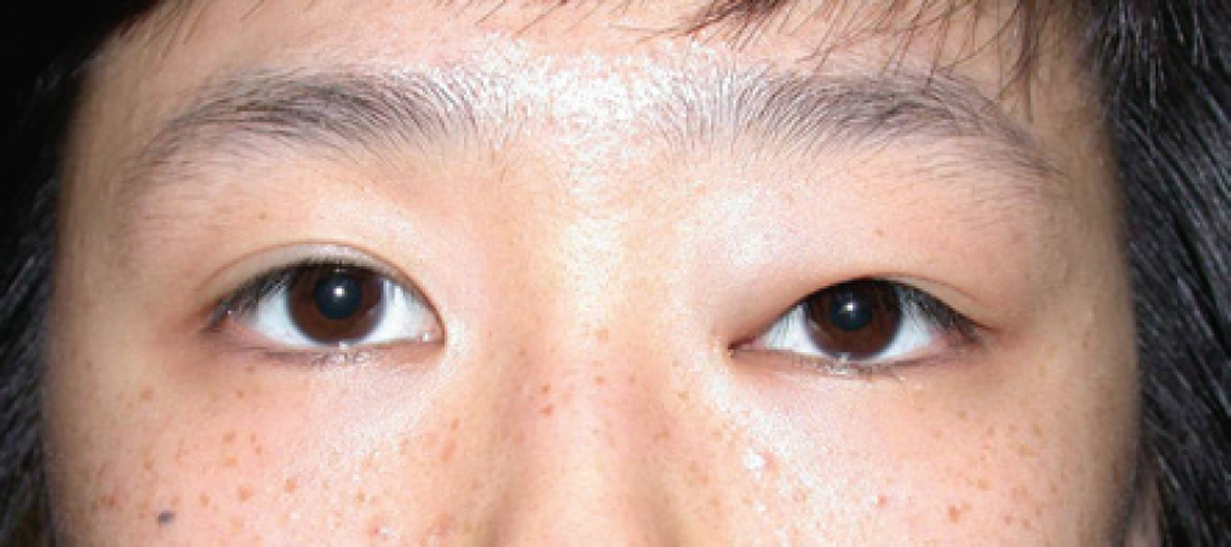
(Case 1) A 17-year-old girl was referred for evaluation of the left upper eyelid swelling which had developed 6 months ago. On initial examination, a 1.5 cm sized ovoid and nontender mass was palpated in the medial aspect of the left orbit. CT scan and MR imaging of the orbit showed a non-calcified, well-circumscribed homogenous soft tissue mass, which was uniformly enhanced and did not invade the adjacent tissue. Excisional biopsy of the orbital mass was performed. (Case 2) A 30-year-old man presented with left proptosis which had developed 2 months ago and hemorrhage into the upper and lower eyelid which had developed 1 week ago. CT scan and MR imaging showed an heterogeneously enhancing mass, not involving the adjacent tissue in the superior retrobulbar space. Excisional biopsy through a lateral orbitotomy was performed. Histologic evaluation revealed proliferation of spindle cells with pseudovascular spaces and multinucleated giant cells.Immunohistochemical staining for CD34 and vimentin was positive and staining for CD31, smooth muscle actin was negative. A diagnosis of giant cell angiofibroma was made.
CONCLUSIONS
The possibility of giant cell angiofibroma should be considered in the differential diagnosis for an orbital mass without a hemorrhage or with a hemorrhage in the eyelid in adult patients.
Keyword
MeSH Terms
Figure
Reference
-
References
1. Keyserling H, Peterson K, Camacho D, Castillo M. Giant cell angiofibroma of the orbit. AJNR Am J Neuroradiol. 2004; 25:1266–8.2. Dei Tos AP, Seregard S, Calonje E, et al. Giant cell angiofibroma. A distinctive orbital tumor in adults. Am J Surg Pathol. 1995; 19:1286–93.3. Mawn LA, Jordan DR, Nerad J, et al. Giant cell angiofibroma of the eyelids: an unusual presentation of tuberous sclerosis. Ophthalmic Surg Lasers. 1999; 30:320–2.
Article4. Husek K, Vesely K. Extraorbital giant cell angiofibroma. Cesk Patol. 2002; 38:117–20.5. Mikami Y, Shimizu M, Hirokawa M, Manabe T. Extraorbital giant cell angiofibromas. Mod Pathol. 1997; 10:1082–7.6. Fukunaga M, Ushigome S. Giant cell angiofibroma of the mediastinum. Histopathology. 1998; 32:187–9.
Article7. Kintarak S, Natiella J, Aguirre A, Brooks J. Giant cell angiofibroma of the buccal mucosa. Oral Surg Oral Med Oral Pathol Oral Radiol Endod. 1999; 88:707–13.
Article8. Rousseau A, Perez-Ordonez B, Jordan RC. Giant cell angiofibroma of the oral cavity: report of a new location for a rare tumor. Oral Surg Oral Med Oral Pathol Oral Radiol Endod. 1999; 88:581–5.
Article9. Wiebe BM, Gottlieb JO, Holck S. Extraorbital giant cell angiofibroma. Apmis. 1999; 107:695–8.
Article10. Yazici B, Setzen G, Meyer DR, et al. Giant cell angiofibroma of the nasolacrimal duct. Ophthal Plast Reconstr Surg. 2001; 17:202–6.
Article11. Song A, Syed N, Kirby PA, Carter KD. Giant cell angiofi- broma of the ocular adnexae. Arch Ophthalmol. 2005; 123:1438–43.12. deSousa JL, Meligonis G, Malhotra R. Giant cell angiofibroma of the orbit with periosteal adherence. Clin Experiment Ophthalmol. 2006; 34:886–8.
Article13. Ereno C, Lopez JI, Perez J, et al. Orbital giant cell angiofibroma. Apmis. 2006; 114:663–5.
Article14. Farmer JP, Lamba M, McDonald H, Commons AS. Orbital giant cell angiofibroma: immuno-histochemistry and differential diagnosis. Can J Ophthalmol. 2006; 41:216–20.
Article15. Qian YW, Malliah R, Lee HJ, et al. A t(12;17) in an extraorbital giant cell angiofibroma. Cancer Genet Cytogenet. 2006; 165:157–60.
Article16. Hayashi N, Borodic G, Karesh JW, et al. Giant cell angiofibroma of the orbit and eyelid. Ophthalmology. 1999; 106:1223–9.
Article17. Guillou L, Gebhard S, Coindre JM. Orbital and extraorbital giant cell angiofibroma: a giant cell-rich variant of solitary fibrous tumor? Clinicopathologic and immunohistochemical analysis of a series in favor of a unifying concept. Am J Surg Pathol. 2000; 24:971–9.18. Mestak J, Urban K, Ondrejka P. Giant cell angiofibroma of the lower eyelids. Cesk Slov Oftalmol. 2002; 58:393–5.19. Font RL, Hidayat AA. Fibrous histiocytoma of the orbit. A clinicopathologic study of 150 cases. Hum Pathol. 1982; 13:199–209.20. Thomas R, Banerjee SS, Eyden BP, et al. A study of four cases of extra-orbital giant cell angiofibroma with documen- tation of some unusual features. Histopathology. 2001; 39:390–6.21. Sonobe H, Iwata J, Komatsu T, et al. A giant cell angiofi- broma involving 6q. Cancer Genet Cytogenet. 2000; 116:47–9.22. Tanaka A, Mihara F, Yoshiura T, et al. Differentiation of cavernous hemangioma from schwannoma of the orbit: a dynamic MRI study. AJR Am J Roentgenol. 2004; 183:1799–804.
- Full Text Links
- Actions
-
Cited
- CITED
-
- Close
- Share
- Similar articles
-
- Primary Angiofibroma Developed in the Orbit
- A Solitary Fibrous Tumor with Giant Cells in the Lacrimal Gland: A Case Study
- Tuberous Sclerosis Occuring through 3 Generations
- A Case of Extranasopharyngeal Angiofibroma Arising from the Inferior Turbinate Removed without Pre-Operative Embolization
- Two Cases of Giant Cell Deficient Giant Cell Tumor of Tendon Sheath


