Anatomic Characteristics of Pronator Quadratus Muscle: A Cadaver Study
- Affiliations
-
- 1Department of Physical Medicine and Rehabilitation, Korea University College of Medicine, Seoul, Korea. rmkdh@korea.ac.kr
- 2Department of Anatomy, Korea University College of Medicine, Seoul, Korea.
- 3Practical Anatomy Center, Korea University College of Medicine, Seoul, Korea.
- KMID: 2327612
- DOI: http://doi.org/10.5535/arm.2016.40.3.496
Abstract
OBJECTIVE
To identify the anatomic characteristics of the pronator quadratus (PQ) muscle and the entry zone (EZ) of the anterior interosseous nerve (AIN) to this muscle by means of cadaver dissection.
METHODS
We examined the PQ muscle and AIN in 20 forearms from 10 fresh cadavers. After identifying the PQ muscle and the EZ of the AIN, we measured the distances from the midpoint (MidP) of the PQ muscle and EZ to the vertical line passing the tip of the ulnar styloid process (MidP_X and EZ_X, respectively) and to the medial border of the ulna (MidP_Y and EZ_Y, respectively). Forearm length (FL) and wrist width (WW) were also measured, and the ratios of MidP and EZ to FL and of MidP and EZ to WW were calculated.
RESULTS
The MidP was found to be 3.0 cm proximal to the ulnar styloid process or distal 13% of the FL and 2.0 cm lateral to the medial border of the ulna or ulnar 40% side of the WW, which was similar to the location of EZ. The results reveal a more distal site than was reported in previous studies.
CONCLUSION
We suggest that the proper site for needle insertion and motor point block of the PQ muscle is 3 cm proximal to the ulnar styloid process or distal 13% of the FL and 2 cm lateral to the medial border of the ulna or ulnar 40% side of the WW.
Keyword
Figure
-
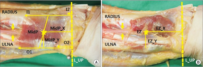
Fig. 1 The origin and insertion site of the pronator quadratus (PQ) muscle was identified (A). The anterior interosseous nerve (AIN) was identified and branches to the PQ muscle were observed (B). L_UP, the vertical line passing the tip of ulnar styloid process; O1, the proximal origin site of the PQ muscle; O2, the distal origin site of the PQ muscle; I1, the proximal insertion site of the PQ muscle; I2, the distal insertion site of the PQ muscle; MidP, the midpoint of the PQ muscle; MidP_X, the distance from MidP to L_UP; MidP_Y, the distance from MidP to the medial border of the ulna; EZ, the entry zone of AIN to the PQ muscle; EZ_X, the distance from EZ to L_UP; EZ_Y, the distance from EZ to the medial border of the ulna.
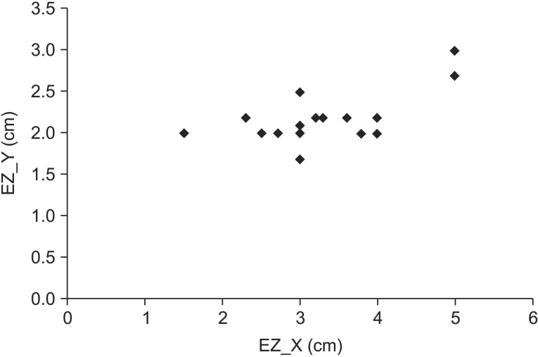
Fig. 2 Plot of the locations of entry zone (EZ) in the pronator quadratus muscle. The x axis is the distance from the vertical line passing the ulnar styloid process to the EZ, and the y axis is the distance from the medial aspect of the ulna to the EZ.
Reference
-
1. Gray H, Standring S, Ellis H, Berkovitz BK. Gray's anatomy: the anatomical basis of clinical practice. 39th ed. Edinburgh: Elsevier Churchill Livingstone;2005.2. Rousseaux M, Kozlowski O, Froger J. Efficacy of botulinum toxin A in upper limb function of hemiplegic patients. J Neurol. 2002; 249:76–84. PMID: 11954872.
Article3. Moore KL, Dalley AF, Agur AM. Clinically oriented anatomy. 7th ed. Philadelphia: Lippincott Williams & Wilkins;2014.4. Lee HJ, DeLisa JA. Manual of nerve conduction study and surface anatomy for needle electromyography. 4th ed. Philadelphia: Lippincott Williams & Wilkins;2005.5. Perotto AO, Delagi EF, Iazzetti J, Morrison D. Anatomical guide for the electromyographer: the limbs and trunk. 4th ed. Springfield: Charles C Thomas Publisher;2005.6. Seror P. Anterior interosseous nerve lesions. Clinical and electrophysiological features. J Bone Joint Surg Br. 1996; 78:238–241. PMID: 8666633.7. Jenkins DB. Hollinshead's functional anatomy of the limbs and back. 9th ed. St. Louis: Saunders;2009.8. Liu J, Lau HK, Min WX, Pereira BP, Kumar VP, Pho RW. Contractile characteristics on electrical stimulation of muscle with multiple motor points. An in vivo study in rabbits. Clin Orthop Relat Res. 1995; 313:231–238. PMID: 7641486.9. Safwat MD, Abdel-Meguid EM. Distribution of terminal nerve entry points to the flexor and extensor groups of forearm muscles: an anatomical study. Folia Morphol (Warsz). 2007; 66:83–93. PMID: 17594664.10. Svizenska I, Cizmar I, Visna P. An anatomical study of the anterior interosseous nerve and its innervation of the pronator quadratus muscle. J Hand Surg Br. 2005; 30:635–637. PMID: 16115708.11. Sakamoto K, Nasu H, Nimura A, Hamada J, Akita K. An anatomic study of the structure and innervation of the pronator quadratus muscle. Anat Sci Int. 2015; 90:82–88. PMID: 24728963.
Article12. Oh SJ. Clinical electromyography: nerve conduction studies. 2nd ed. Philadelphia: Lippincott Williams & Wilkins;1993.13. Shafshak TS, El-Hinawy YM. The anterior interosseous nerve latency in the diagnosis of severe carpal tunnel syndrome with unobtainable median nerve distal conduction. Arch Phys Med Rehabil. 1995; 76:471–475. PMID: 7741621.
Article14. Foley BS, Roslonski ET, Buschbacher RM. Reference values for median nerve conduction to the pronator quadratus. Arch Phys Med Rehabil. 2006; 87:88–91. PMID: 16401444.
Article15. Dumitru D, Amato AA, Zwarts MJ. Electrodiagnostic medicine. 2nd ed. Philadelphia: Hanley & Belfus;2002.16. Lin DL, Lenhart MK, Farber GL. Anatomy of the anterior interosseous innervation of the pronator quadratus: evaluation of structures at risk in the single dorsal incision wrist denervation technique. J Hand Surg Am. 2006; 31:904–907. PMID: 16843148.
Article17. Alves N, Candido PL, Frazao R. Innervation of the pronator teres muscle. Int J Morphol. 2004; 22:237–240.
Article
- Full Text Links
-
- Actions
-
Cited
- CITED
-
- Close
- Share
-
- Similar articles
-
- Anatomical Study of the Pronator Quadratus Muscle and Comparison to Fracture Sites of the Distal Radius
- The Fate of Pronator Quadratus Muscle after Volar Locking Plating of Unstable Distal Radius Fractures
- Treatment of Scaphoid Nonunions Using a Pronator Quadratus Pedicled Bone Graft
- A Case of Extrahepatic Metastasis of Hepatocellular Carcinoma to the Pronator Quadratus Muscle of Right Wrist
- Ultrasonographic Assessment of the Pronator Quadratus Muscle after Surgical Treatment for Distal Radius Fractures
