An Unreported Type of Coronary Artery Anomaly in Congenitally Corrected Transposition of Great Arteries
- Affiliations
-
- 1Department of Radiology, Medical Research Institute, Pusan National University Hospital, Busan, Korea. jw@pusan.ac.kr
- 2Department of Internal Medicine, Medical Research Institute, Pusan National University Hospital, Busan, Korea.
- KMID: 2327369
- DOI: http://doi.org/10.3348/jksr.2016.75.1.62
Abstract
- Coronary artery variations are associated anomalies in 45% of congenitally corrected transposition of the great arteries (ccTGA) cases, and it is important to detect any coronary artery anomalies before cardiac surgery. We report a case of a 51-year-old woman with ccTGA and an unreported type of coronary artery anomaly.
MeSH Terms
Figure
-
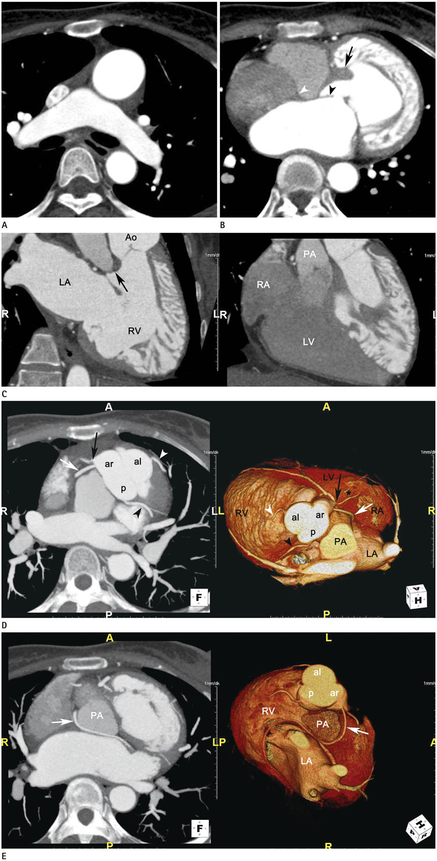
Fig. 1 An unreported type of coronary artery anomaly in congenitally corrected transposition of great arteries. A. Axial CT image shows the aorta anterior to and left of the pulmonary trunk, suggestive of levo-transposition of the great vessels. B. Axial CT image shows that the left-sided AV valve (black arrowhead) lies closer to the apex than does the right-sided AV valve (white arrowhead). A moderator band (arrow) and prominent trabeculation are seen in the left-sided ventricle. C. The three-chamber view of the left-sided ventricle shows a tricuspid-aortic fibrous discontinuity (black arrow) caused by the presence of the right ventricular infundibulum (left). The three-chamber view of the right-sided ventricle shows mitral-pulmonary fibrous continuity (right). Ao = aorta, AV = atrioventricular, CT = computed tomography, LA = left atrium, LV = morphological left ventricle, PA = pulmonary artery, RA = right atrium, RV = morphological right ventricle D. Oblique axial MIP and 3D VR images show coronary arteries arising from three separate coronary ostia. A stair-step artifact (*) resulting from the irregular heart rhythm in the proximal segment of the circumflex artery arising from the anterior right sinus is also seen. The aortic valve has three cusps, which are located to the left anteriorly, right anteriorly, and posteriorly. The right ventricular branch (white arrowhead), which runs along the wall on the morphological right ventricle, originates from the anterior left sinus. The circumflex and anterior descending arteries arise from one ostium (black arrow) of the anterior right sinus. The right ventricular artery (white arrow) originates from the other ostium of the anterior right sinus. Another right ventricular branch (black arrowhead) originates from the posterior sinus. E. Axial MIP and 3D VR images show the right ventricular artery arising from the anterior right sinus traveling along the left AV groove with a retropulmonary course (white arrow). al = anterior left sinus, ar = anterior right sinus, CT = computed tomography, LA = left atrium, LV = morphological left ventricle, MIP = maximum intensity projection, p = posterior sinus, PA = pulmonary artery, RA = right atrium, RV = morphological right ventricle, 3D VR = 3D dimensional volume-rendered
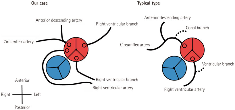
Fig. 2 Diagrams of the coronary anatomy showing our case and typical ccTGA. Dashed lines represent variably present branches. ccTGA = congenitally corrected transposition of the great arteries
Reference
-
1. Wallis GA, Debich-Spicer D, Anderson RH. Congenitally corrected transposition. Orphanet J Rare Dis. 2011; 6:22.2. Warnes CA. Transposition of the great arteries. Circulation. 2006; 114:2699–2709.3. Huang SC, Chiu IS, Lee ML, Wu CS, Chiu HH, Chang CI, et al. Coronary artery anatomy in anatomically corrected malposition of the great arteries and their surgical implications. Eur J Cardiothorac Surg. 2011; 39:705–710.4. Ismat FA, Baldwin HS, Karl TR, Weinberg PM. Coronary anatomy in congenitally corrected transposition of the great arteries. Int J Cardiol. 2002; 86:207–216.5. Friedberg DZ, Nadas AS. Clinical profile of patients with congenital corrected transposition of the great arteries. A study of 60 cases. N Engl J Med. 1970; 282:1053–1059.6. Malhotra S, Patel RN, Mandawat M. A case of congenitally corrected transposition of the great arteries with rare but life-threatening ventricular tachycardia and a coincidental single coronary ostium. J Invasive Cardiol. 2007; 19:E139–E141.7. Karl TR. The role of the fontan operation in the treatment of congenitally corrected transposition of the great arteries. Ann Pediatr Cardiol. 2011; 4:103–110.8. Sithamparanathan S, Padley SP, Rubens MB, Gatzoulis MA, Ho SY, Nicol ED. Great vessel and coronary artery anatomy in transposition and other coronary anomalies: a universal descriptive and alphanumerical sequential classification. JACC Cardiovasc Imaging. 2013; 6:624–630.9. Kantarci M, Koplay M, Bayraktutan U, Gundogdu F, Ceviz N. Congenitally corrected transposition of the great arteries: MDCT angiography findings and interpretation of complex coronary anatomy. Int J Cardiovasc Imaging. 2007; 23:405–410.10. Chiu IS, Wu SJ, Chen SJ, Wang JK, Wu MH, Lue HC. Sequential diagnosis of coronary arterial anatomy in congenitally corrected transposition of the great arteries. Ann Thorac Surg. 2003; 75:422–429.
- Full Text Links
-
- Actions
-
Cited
- CITED
-
- Close
- Share
-
- Similar articles
-
- Transposition of the Great Arteries: Historical Background
- A Case Report of Congenitally Corrected Transposition of Great Arteries: Morphologic and Functional Evaluation with Cardiac CT
- An Adult Case of Congenitally Corrected Transposition of the Great Arteries Associated with Paroxysmal Atrial Fibrillation and Heart Failure
- Q waves in congenitally corrected transposition of the great arteries
- Coronary Angiography in an Adult Case of lsolated Congenitally Corrected Transposition of the Great Vessels
