J Korean Soc Radiol.
2016 Jul;75(1):30-36. 10.3348/jksr.2016.75.1.30.
Comparative Analysis of Maximum Renal Longitudinal Length with Positional Changes on Ultrasound with Multiplanar Reconstructed MR Image in Korean Adults
- Affiliations
-
- 1Department of Radiology, Chungbuk National University Hospital, Cheongju, Korea. sircircle@hanmail.net
- 2Department of Radiology, College of Medicine and Medical Research Institute, Chungbuk National University, Cheongju, Korea.
- 3Public Health Medical Service, Seoul National University Boramae Medical Center, Seoul, Korea.
- KMID: 2327364
- DOI: http://doi.org/10.3348/jksr.2016.75.1.30
Abstract
- PURPOSE
The purpose of this study was to determine a suitable position in which the measured length on ultrasound is close to the true renal length obtained through a multiplanar reconstructed MR image.
MATERIALS AND METHODS
A total of 33 individuals (males: 15, females: 18) without any underlying renal disease were included in the present study. Renal length was measured as the longest axis at the level of the renal hilum in three positions-supine, lateral decubitus, and prone, respectively. With a 3.0 T MR scanner, 3D eTHRIVE was acquired. Subsequently, the maximum longitudinal length of both the kidneys was measured through multiplanar reconstructed MR images. Paired t-test was used to compare the renal length obtained from ultrasonographic measurement with the length obtained through multiplanar reconstructed MR images.
RESULTS
Our study demonstrated significant difference between sonographic renal length in three positions and renal length through MRI (p < 0.001). However, the longest longitudinal length of right kidney among the measured three values by ultrasound was statistically similar to the renal length measured by reconstructed MR image. Among them, the lateral decubitus position showed the strongest correlation with true renal length (right: 0.887; left: 0.849).
CONCLUSION
We recommend measurement of the maximum renal longitudinal length in all possible positions on ultrasonography. If not allowed, the best measurement is on the lateral decubitus showing the strongest correlation coefficient with true renal length.
Figure
-
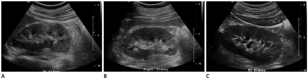
Fig. 1 Ultrasonographic measurement of maximum renal length in supine (10.7 cm) (A), lateral decubitus (10.3 cm) (B), and prone position (10.4 cm) (C) right kidney of a 31-year-old man without any underlying renal disease.
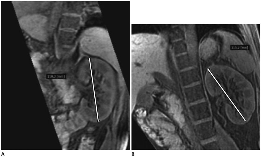
Fig. 2 Maximum renal length measured by multiplanar reconstructed MR image of a 32-year-old man without any underlying renal disease. Right kidney (11.0 cm) (A) and left kidney (11.5 cm) (close-cut image) (B).
Reference
-
1. Kim JH, Kim MJ, Lim SH, Kim J, Lee MJ. Length and volume of morphologically normal kidneys in Korean children: ultrasound measurement and estimation using body size. Korean J Radiol. 2013; 14:677–682.2. Kasper DL, Longo DL, Fauci AS, Braunwald E, Hauser S, Jameson JL. Harrison's principles of internal medicine. 16th ed. New York: McGraw-Hill;2004. p. 1653–1663.3. Lisanti CJ, Oettel DJ, Reiter MJ, Schwope RB. Multiplanar reformations in the measurement of renal length on CT: is it plain which plane to use? AJR Am J Roentgenol. 2015; 205:797–801.4. Edell SL, Kurtz AB, Rifkin MD. Normal renal ultrasound measurements. In : Goldberg BB, Kurtz AB, editors. Atlas of ultrasound measurements. Chicago: Mosby-Year Book;1990. p. 146–160.5. Bakker J, Olree M, Kaatee R, de Lange EE, Moons KG, Beutler JJ, et al. Renal volume measurements: accuracy and repeatability of US compared with that of MR imaging. Radiology. 1999; 211:623–628.6. Michel SC, Forster I, Seifert B, Willi UV, Huisman TA. Renal dimensions measured by ultrasonography in children: variations as a function of the imaging plane and patient position. Eur Radiol. 2004; 14:1508–1512.7. De Sanctis JT, Connolly SA, Bramson RT. Effect of patient position on sonographically measured renal length in neonates, infants, and children. AJR Am J Roentgenol. 1998; 170:1381–1383.8. Alexander MP, Patel TV, Farag YM, Florez A, Rennke HG, Singh AK. Kidney pathological changes in metabolic syndrome: a cross-sectional study. Am J Kidney Dis. 2009; 53:751–759.9. Zhang WX, Zhang ZM, Cao BS, Zhou W. Sonographic measurement of renal size in patients undergoing chronic hemodialysis: correlation with residual renal function. Exp Ther Med. 2014; 7:1259–1264.10. Kim SH. Radiology illustrated: uroradiology. 2nd ed. Berlin: Springer-Verlag;2012. p. 56.11. Emamian SA, Nielsen MB, Pedersen JF, Ytte L. Kidney dimensions at sonography: correlation with age, sex, and habitus in 665 adult volunteers. AJR Am J Roentgenol. 1993; 160:83–86.12. Kang KY, Lee YJ, Park SC, Yang CW, Kim YS, Moon IS, et al. A comparative study of methods of estimating kidney length in kidney transplantation donors. Nephrol Dial Transplant. 2007; 22:2322–2327.13. Bhutani H, Smith V, Rahbari-Oskoui F, Mittal A, Grantham JJ, Torres VE, et al. A comparison of ultrasound and magnetic resonance imaging shows that kidney length predicts chronic kidney disease in autosomal dominant polycystic kidney disease. Kidney Int. 2015; 88:146–151.14. Chapple CR, Steers WD. Practical urology: essential principles and practice: essential principles and practice. London: Springer Science & Business Media;2011. p. 32.15. Sanders RC, Winter TC. Clinical sonography: a practical guide. 4th ed. Philadelphia: Lippincott Williams & Wilkins;2007. p. 123–124.
- Full Text Links
-
- Actions
-
Cited
- CITED
-
- Close
- Share
-
- Similar articles
-
- Anatomic illustrations of Cranial Ultrasound Images in the Neonate: Objective Analysis of the Oblique Sonographic Scans using MRI and a Reconstruction Program
- Utility of Multiplanar Reformation Images of Helical CT in the Evaluation of Pancreatic Diseases
- Applications of Artificial Intelligence in MR Image Acquisition and Reconstruction
- Contrast-Enhanced Three-Dimensional MR Imaging Using a Volumetric Interpolated Breath-hold Examination (VIBE): Clinical Utility in the Evaluation of Renal Tumors
- Hard- and soft-tissue profiles of the midface region in patients with skeletal Class III malocclusion using cone-beam computed tomography multiplanar-reconstructed image analysis
