Degenerative Changes of Adjacent Segment Following Cervical Anterior fusion: Correlation with Preoperative MRI Findings of Adjacent Disc
- Affiliations
-
- 1Department of Orthopedic Surgery, Seoul Paik Hospital, Inje University, Seoul, Korea. hd1404@hanafos.com
Abstract
- STUDY DESIGN: We retrospectively reviewed the preoperative and postoperative radiographs of patients who underwent anterior cervical discectomy and fusion.
OBJECTIVES
We wanted to determine whether the preoperative Magnetic Resonance Imaging (MRI) findings of the levels adjacent to the level of fusion correlated with the postoperative degenerative changes seen on X-ray after anterior cervical discectomy and fusion. SUMMARY OF LITERATURE REVIEW: Anterior cervical fusion causes acceleration of the degenerative changes at the levels below or above the fused segment. These changes may be accelerated if preoperative MRI shows degenerative changes at the levels adjacent to the segment to be fused.
MATERIALS AND METHODS
Twenty-two patients (forty-four adjacent levels) who underwent anterior cervical discectomy and fusion from January 1998 to August 2002 (average follow up: 2 years and 6 months, range: 2 to 4 years) were enrolled in this study. Preoperatively, all the patients had no degenerative changes at adjacent levels on the plain radiographs, but they had at least one adjacent level with degenerative findings on MRI. The patients were grouped according to the findings of the adjacent levels seen on MRI: low signal changes on the T2 weighted image (group A), disc bulging on the sagittal and axial images (group B), annular tear seen on the axial image (group C), osteophyte formation (group D), and no abnormalities (group E).
RESULTS
Out of 44 cases of 22 patients, 14 cases (31.8%) showed degenerative changes. 2 out of 7 in group A, 6 out of 11 in group B, 3 out of 4 in group C, 2 out of 3 in group D and 1 out of 19 in group E showed degenerative changes on X-rays at the final follow up.
CONCLUSION
Our findings suggest that abnormalities on the levels adjacent to the level to be fused, as seen on preoperative MRI, predispose these levels to degenerative changes postoperatively.
MeSH Terms
Figure
-

Fig. 1. Degenerative findings of adjacent levels seen on MRI (A) Group A. Disc herniation at C3-4 with low signal change seen at lower adjacent level, C4-5, on T2 weighted image. (B) Group B. Disc herniation at C4-5 with disc bulging at lower adjacent level, C5-6. (C) Group C. Annular tear shown on axial image. (D) Group D. Disc herniation at C5-6 with osteophyte formation at upper adjacent level, C4-5.
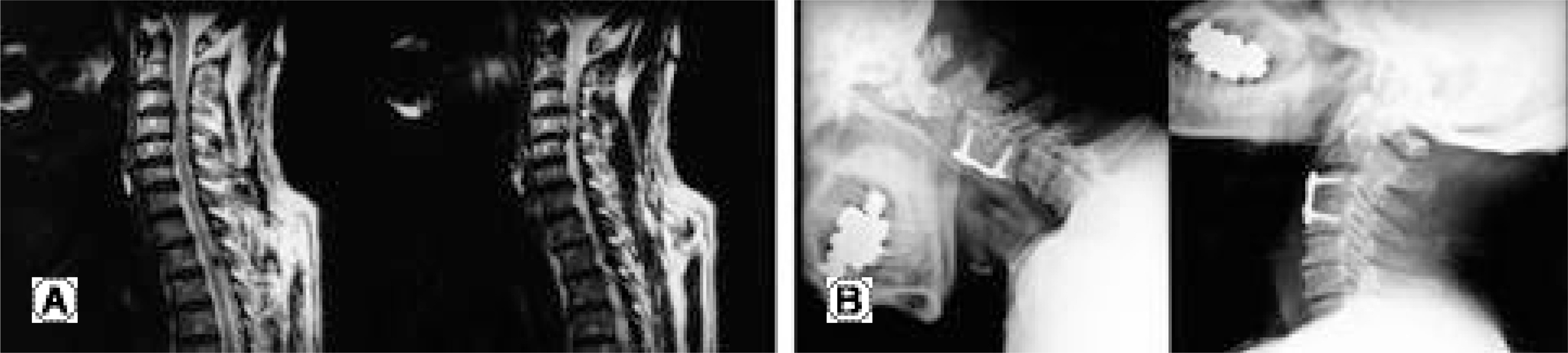
Fig. 2. This 67-year old male patient underwent anterior cervical discectomy and fusion for herniated intervertebral disc at C3-4. Pre-operative X-ray showed no degenerative changes at adjacent levels of C3-4 (A) But preoperative T2 weighted sagittal MR image showed protrusion of the disc at C3-4. Adjacent to this level, C4-5 disc showed bulging, which was categorized as group B. Disc at C2-3 showed no degenerative changes and subsequently were categorized as group E. (B) Twenty-seven months after anterior cervical discectomy and fusion was done using autogenous iliac bone graft and ORION plate, X-ray shows that degenerative changes have occurred at lower adjacent level, C4-5, with narrowing of the disc space and spur formation. The upper adjacent level, C2-3, showed no degenerative changes.
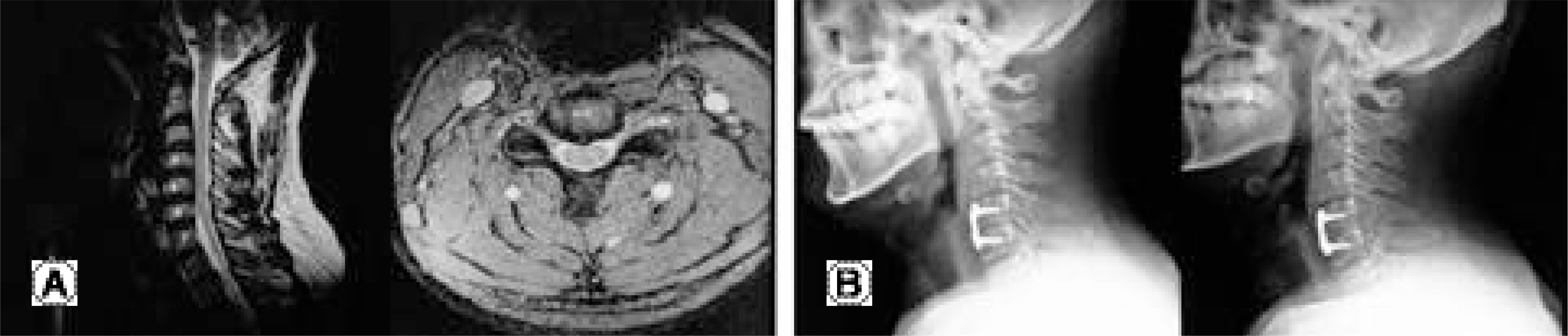
Fig. 3. This 44-year old male patient underwent anterior cervical discectomy and fusion for herniated intervertebral disc at C5-6. Pre-operative X-ray showed no degenerative changes at C4-5 or at C6-7 (A) But preoperative T2 weighted sagittal MR image showed protrusion of the disc at C5-6. Adjacent to this level, C4-5 disc showed annular tear on T2 weighted axial image, which was categorized as group C. Disc at C6-7 showed no degenerative changes and subsequently were categorized as group E. (B) Twenty-six months after anterior cervical discectomy and fusion was done using autogenous iliac bone graft and ORION plate, X-ray shows that degenerative changes have occurred at upper adjacent level, C4-5, with narrowing of the disc space and spur formation. The lower adjacent level, C6-7, showed no degenerative changes.
Reference
-
1). Robinson RA and Smith GW. Anterolateral cervical disc removing and interbody fusion for cervical disc syndrome. Bull John Hopkins Hospital. 1955; 96:223–224.2). Braunstein EM, Hunter LY, Bailey RW. Long term radiographic changes following anterior cervical fusion. Clin Radiol. 1980; 31:201–203.
Article3). Gore DR, Sepic SB. Anterior cervical fusion for degenerated or protruded discs: A review of one hundred forty-six patients. Spine. 1984; 9:667–671.4). Gore DR, Sepic SB. Anterior discectomy and fusion for painful cervical disc disease. A report of 50 patients with an average follow-up of 21 years. Spine. 1998; 23:2047–2051.5). Shinomiya K, Okamoto A, Kamikozuru M, Furuya K, Yamaur I. An analysis of failures in primary cervical anterior spinal cord decompression and fusion. J Spinal Disord. 1993; 6:277–288.
Article6). Wu W, Thuomas KA, Hedlund R, Leszniewski W, Vavruch L. Degenerative changes following anterior cervical discectomy and fusion evaluated by fast spin-echo MR imaging. Acta Radiol. 1996; 37:614–617.
Article7). Yonenobu K, Okada K, Fuji T, Fujiwara K, Yamashita K, Ono K. Causes of neurologic deterioration following surgical treatment of cervical myelopathy. Spine. 1986; 11:818–823.
Article8). Goto S, Mochizuki M, Kita T, et al. Anterior surgery in four consecutive technical phases for cervical spondylotic myelopathy. Spine. 1993; 18:1968–1973.
Article9). Bohlman HH, Emery SE, Goodfellow DB, Jones PK. Robinson anterior cervical discectomy and arthrodesis for cervical radiculopathy: Long-term follow-up of one hundred and twenty-two patients. J Bone Joint Surg Am. 1993; 75:1298–1307.
Article10). Robinson RA, Walker AE, Ferlic DC, Wiecking DK. The results of anterior interbody fusion of the cervical spine. J Bone Joint Surg. 1962; 44:1569–1587.
Article11). Ahn JS, Lee JK, Yang JY, Lee HH. Change of the lordosis on cervical spine after anterior interbody fusion with antogenous iliac strut boen graft. J Kor Spine Surg. 2001; 8:468–474.12). Park HJ, Rha JH, Yoon YS. Anterior cervical interbody fusion using cervical locking plate (CSLP). J Kor Orthop Assoc. 1996; 31:52–57.13). Harris RI, Wiley JJ. Acquired spondylosis as a sequel to spine fusion. J Bone Joint Surg Am. 1963; 45:1159–1170.14). Baba H, Furusawa N, Imura S, Kawahara N, Tsuchiya H, Tomita K. Late radiographic findings after anterior cervical fusion for spondylotic myeloradiculopathy. Spine. 1993; 18:2167–2173.
Article15). Gore DR, Gardner GM, Sepic SB, Murray MP. Roentgenographic findings following anterior cervical fusion. Skeletal Radiol. 1986; 15:556–9.
Article16). Hunter LY, Braunstein EM, Bailey RW. Radiographic changes following anterior cervical fusion. Spine. 1980; 5:399–401.
Article17). Gregorius FK, Estrin T, Crandall PH. Cervical spondylotic radiculopathy and myelopathy. A long-term follow-up study. Arch Neurol. 1976; 33:618–625.18). Jackson RP, McManus AC. Radiographic analysis of sagittal plane alignment and balance in standing volunteers and patients with low back pain matched for age, sex and size; a prospective controled clinical study. Spine. 1994; 19:1611–1618.19). Oda I, Cunningham BW, Buckley RA, et al. Does spinal kyphotic deformity influence the biomechanical characteristics of the adjacent motion segments An in vivo animal model. Spine. 1999; 24:2139–2146.20). Katsuura A, Hukuda S, Saruhashi Y, Mori K. Kyphotic malalignment after anterior cervical fusion in one of the factors promoting the degenrative process in adjacent intervetabral levels. Eur spine J. 2001; 10:320–324.21). Dohler JR, Kahn MR, Hughes SP. Instability of the cervical spine after anterior interbody fusion. A study on its incidence and clinical significance in 21 patients. Acta Orthop trauma Surg. 1985; 104:247–250.22). Fuller DA, Kirkpatrick JS, Emery SE, Wilber RG, Davy DT. A kinematic study after cervical spine before and after segmental arthrodesis. Spine. 1998; 23:1649–1656.23). Cloward RB. The anterior approach for removal of ruptured cervical disks. J Neurosurg. 1958; 15:602–617.
Article24). Dvorak J, Froehlich D, Penning L, Baumgartner H, Panjabi MM. Functional radiographic diagnosis of the cervical spine: Flexion/Extension. Spine. 1988; 13:748–755.
Article
- Full Text Links
-
- Actions
-
Cited
- CITED
-
- Close
- Share
-
- Similar articles
-
- Changes of Adjacent Segment in Anterior Cervical Fusion
- New Classification for Clinically Symptomatic Adjacent Segment Pathology in Cervical Disc Disease
- Degenerative Changes of Adjacent Segment after Anterior Cervical Discectomy and Fusion
- Risk Factors for “Adjacent-Level Ossification Development” Other Than Short Plate-to-Disc Distance and Clinical Implications for Adjacent-Segment Pathology
- Radiologic Changes of Operated and Adjacent Segments after Anterior Cervical Microforaminotomy
