Analysis of Bone Mineral Density in Multiple Myeloma: A Comparison of Bone Mineral Density with Plain Radiography, Magnetic Resonance Imaging, and Clinical Staging
- Affiliations
-
- 1Department of Radiology, Chungnam National University Hospital, Daejeon, Korea. stkwon@cnu.ac.kr
- 2Department of Internal Medicine, Chungnam National University Hospital, Daejeon, Korea.
Abstract
- PURPOSE
To analyze the bone mineral density (BMD) in multiple myeloma (MM) and to compare BMD with plain radiography, MRI and clinical stage.
MATERIALS AND METHODS
We reviewed 59 patients with MM and an age- and sex-matched control group, with measured BMD. The L-spine and femoral neck (FN) BMD were measured by dual-energy X-ray absorptiometry. Lateral plain radiographs of the L-spine were graded as 3 stages using the modified Saville index. Four bone marrow patterns were classified on sagittal T1- and T2-weighted magnetic resonance images of the L-spine. BMD in the MM and control group were analyzed. BMD in MM was compared with the modified Saville index, bone marrow patterns on MRI, and clinical stages.
RESULTS
In MM, spine BMD was reduced and the difference between spine and FN BMD was larger than the control group (p < 0.001). The modified Saville index was negatively correlated with spine T scores (p < 0.01). The spine BMD in normal marrow pattern on the MRI was the most reduced. There was no statistical correlation between BMD and clinical stage.
CONCLUSION
In MM, spine BMD was significantly reduced and the difference between spine and FN BMD was larger than the control group. The modified Saville index was significantly correlated with spine BMD in MM.
MeSH Terms
Figure
-

Fig. 1 BMD in MM and control group. A. Spine BMD was significantly reduced in MM patients compared to the control group (p < 0.001). B. The difference between spine and femoral neck BMD in MM was larger than that in the control group (p < 0.001). Note.-BMD = bone mineral density, MM = multiple myeloma
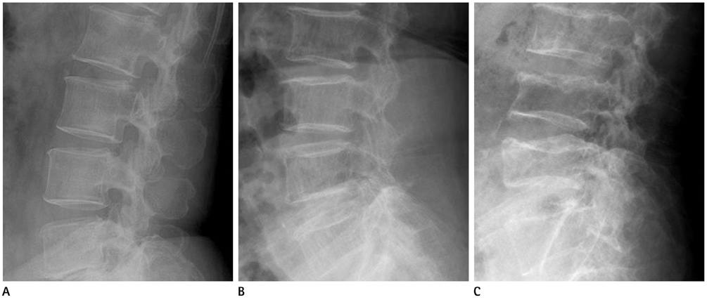
Fig. 2 Modified Saville index. A. Lateral plain radiograph of L-spine shows minimal decreased bone density indicating modified Saville index grade 1. B. More obvious vertical striation with concave endplates reveals modified Saville index grade 2. C. Severe diminished bone density and a more collapsed vertebral body indicates modified Saville index grade 3.
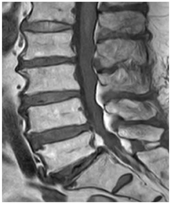
Fig. 3 Normal marrow pattern on magnetic resonance images. Bone marrow shows hyperintense signal compared with the disc on T1-weighted images.
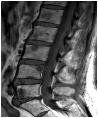
Fig. 4 Multiple focal pattern on magnetic resonance images. Multifocal T1-weighted hypointense lesions are detected in the L-spine.
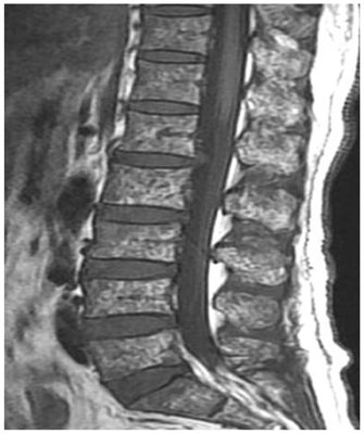
Fig. 5 Salt-and-pepper pattern. The bone marrow shows a very inhomogeneous patchy signal on T1-weighted images.
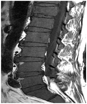
Fig. 6 Diffuse infiltrative pattern. Homogenous hypointense change which is similar or lower than disc signal intensity on T1-weighted images is shown in the spine.
Reference
-
1. Mounach A, Abayi DA, Ghazi M, Ghozlani I, Nouijai A, Achemlal L, et al. Discordance between hip and spine bone mineral density measurement using DXA: prevalence and risk factors. Semin Arthritis Rheum. 2009. 38:467–471.2. Saville PD. A quantitative approach to simple radiographic diagnosis of osteoporosis: its application to the osteoporosis of rheumatoid arthritis. Arthritis Rheum. 1967. 10:416–422.3. Mariette X, Zagdanski AM, Guermazi A, Bergot C, Arnould A, Frija J, et al. Prognostic value of vertebral lesions detected by magnetic resonance imaging in patients with stage I multiple myeloma. Br J Haematol. 1999. 104:723–729.4. Baur A, Stäbler A, Bartl R, Lamerz R, Scheidler J, Reiser M. MRI gadolinium enhancement of bone marrow: age-related changes in normals and in diffuse neoplastic infiltration. Skeletal Radiol. 1997. 26:414–418.5. Durie BG, Salmon SE. A clinical staging system for multiple myeloma. Correlation of measured myeloma cell mass with presenting clinical features, response to treatment, and survival. Cancer. 1975. 36:842–854.6. Greipp PR, San Miguel J, Durie BG, Crowley JJ, Barlogie B, Bladé J, et al. International staging system for multiple myeloma. J Clin Oncol. 2005. 23:3412–3420.7. Abildgaard N, Brixen K, Eriksen EF, Kristensen JE, Nielsen JL, Heickendorff L. Sequential analysis of biochemical markers of bone resorption and bone densitometry in multiple myeloma. Haematologica. 2004. 89:567–577.8. Lütje S, de Rooy JW, Croockewit S, Koedam E, Oyen WJ, Raymakers RA. Role of radiography, MRI and FDG-PET/CT in diagnosing, staging and therapeutical evaluation of patients with multiple myeloma. Ann Hematol. 2009. 88:1161–1168.9. Dhodapkar MV, Weinstein R, Tricot G, Jagannath S, Parfitt AM, Manolagas SC, et al. Biologic and therapeutic determinants of bone mineral density in multiple myeloma. Leuk Lymphoma. 1998. 32:121–127.10. Johnston CC Jr, Slemenda CW, Melton LJ 3rd. Clinical use of bone densitometry. N Engl J Med. 1991. 324:1105–1109.11. Mariette X, Khalifa P, Ravaud P, Frija J, Laval-Jeantet M, Chastang C, et al. Bone densitometry in patients with multiple myeloma. Am J Med. 1992. 93:595–598.12. Karsh J. Diagnostic challenges in osteoporosis. Indications for bone densitometry and establishing secondary causes. Can Fam Physician. 2001. 47:1244–1250.13. Mariette X, Bergot C, Ravaud P, Roux C, Laval-Jeantet M, Brouet JC, et al. Evolution of bone densitometry in patients with myeloma treated with conventional or intensive therapy. Cancer. 1995. 76:1559–1563.14. Abildgaard N, Brixen K, Kristensen JE, Vejlgaard T, Charles P, Nielsen JL. Assessment of bone involvement in patients with multiple myeloma using bone densitometry. Eur J Haematol. 1996. 57:370–376.15. Hanrahan CJ, Christensen CR, Crim JR. Current concepts in the evaluation of multiple myeloma with MR imaging and FDG PET/CT. Radiographics. 2010. 30:127–142.16. Fechtner K, Hillengass J, Delorme S, Heiss C, Neben K, Goldschmidt H, et al. Staging monoclonal plasma cell disease: comparison of the Durie-Salmon and the Durie-Salmon PLUS staging systems. Radiology. 2010. 257:195–204.17. Baur-Melnyk A, Buhmann S, Dürr HR, Reiser M. Role of MRI for the diagnosis and prognosis of multiple myeloma. Eur J Radiol. 2005. 55:56–63.18. Moulopoulos LA, Varma DG, Dimopoulos MA, Leeds NE, Kim EE, Johnston DA, et al. Multiple myeloma: spinal MR imaging in patients with untreated newly diagnosed disease. Radiology. 1992. 185:833–840.
- Full Text Links
-
- Actions
-
Cited
- CITED
-
- Close
- Share
-
- Similar articles
-
- The relationship of maturation value of vaginal epithelium and bone mineral density in postmenopausal women
- Measurement of bone mineral density in Korean newborns by dual photon absorptiometry
- Effects of Body Composition, Leptin, and Adiponectin on Bone Mineral Density in Prepubertal Girls
- Diagnostic Value of the Bone Mineral Densitometry in the Metastatic Prostatic Cancer
- Osteoporosis
