Comparison of Different 3.0 T Magnetic Resonance Sequences for Lumbosacral Plexus and Its Branches: Preliminary Study
- Affiliations
-
- 1Department of Radiology, Seoul National University Bundang Hospital, Seongnam, Korea. joonwoo2@gmail.com
Abstract
- PURPOSE
To prospectively evaluate four magnetic resonance sequences [ProSet, fluid attenuation inversion recovery (FLAIR), balanced turbo field echo (B-TFE), T2 Drive] for the lumbosacral plexus and its branches.
MATERIALS AND METHODS
Ten healthy volunteers who underwent four MRI sequences on lumbosacral area were evaluated for image quality (1 to 5; 1 = poor, 5 = excellent), the number of visualized bilateral spinal nerves from L2 to S1, the overlapping vessels obscuring the plexus (1 = many, 2 = some, 3 = few), and image quality defining spinal nerves (0 = nonvisualized, 1 = poor, 2 = moderate, 3 = good).
RESULTS
The ProSet (mean = 4.2, range 3-5) and B-TFE (mean = 3.7, range 3-5) showed better image quality than others. The number of visualized spinal nerves was the largest on ProSet image (mean = 9.2, range 8-10). FLAIR (mean = 2.1, range 1-3) and T2 Drive sequences (mean = 2.1, range 1-3) discriminated the nerves well from the vessels. The main branches of the lumbosacral plexus were well visualized on both ProSet (mean = 2.9, range 2-3) and FLAIR images (mean = 2.6, range 1-3). All of these were statistically significant.
CONCLUSION
ProSet is the best sequence in the evaluation of the lumbosacral plexus and its major branches while FLAIR can be a complementary sequence for the evaluation of nerves overlapping vascular structures.
MeSH Terms
Figure
-
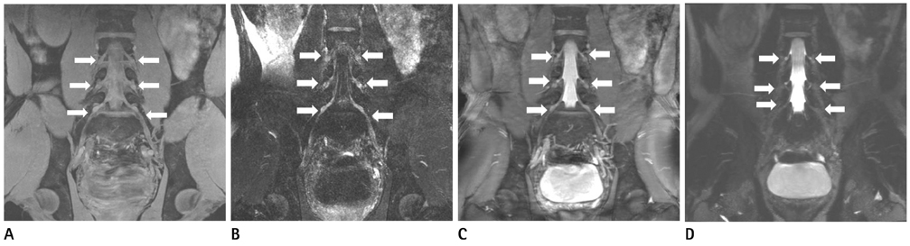
Fig. 1 Coronal reformatted images of four different sequences of 29-year-old healthy male. Spinal nerves and dorsal root ganglia (arrows) are seen on images of four different sequences, and those are best visualized on ProSet image. The image quality for spinal nerve visualization on ProSet image (A) and B-TFE image (C) are better than those on FLAIR image (B) and T2 Drive image (D). Note.-B-TFE = balanced turbo field echo, FLAIR = fluid attenuation inversion recovery
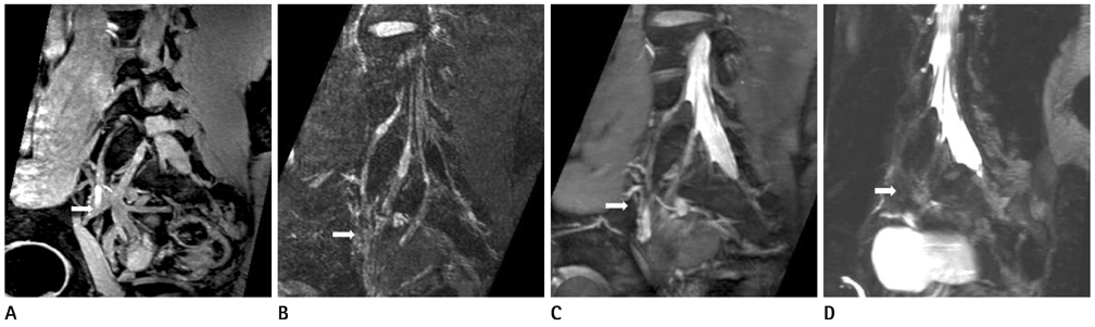
Fig. 2 Oblique sagittal reformatted images of four different sequences of 29-year-old healthy male. Vessels could pass close by the pathway of spinal nerves. Some portions of the nerves are obscured by the vessels on ProSet image (A), B-TFE image (C) and T2 Drive image (D) (arrows on A, C and D). However, the nerves on FLAIR image (B) are well-discriminated from the adjacent vessels (arrow on B). Note.-B-TFE = balanced turbo field echo, FLAIR = fluid attenuation inversion recovery

Fig. 3 Oblique coronal reformatted images of four different sequences of 32-year-old healthy male. Tibial nerve (arrows) and sciatic nerve (arrowheads) are relatively well demonstrated on ProSet and FLAIR images (A: ProSet image, B: FLAIR image, C: B-TFE image, D: T2 Drive image). Note.-B-TFE = balanced turbo field echo, FLAIR = fluid attenuation inversion recovery

Fig. 4 Oblique sagittal reformatted images of four different sequences of 30-year-old healthy male. FLAIR image is the best sequence to discriminate femoral nerve (arrows) from surrounding structures (A: ProSet image, B: FLAIR image, C: B-TFE image, D: T2 Drive image). Note.-B-TFE = balanced turbo field echo, FLAIR = fluid attenuation inversion recovery

Fig. 5 Oblique sagittal reformatted images of four different sequences of 28-year-old healthy male. On ProSet sequence, obturator nerve (arrows) is well-demarcated by surrounding low signal fat plane as thin and intermediate signal structure. On T2 Drive image, the obturator nerve is not distinguished from surrounding fat plane (A: ProSet image, B: FLAIR image, C: B-TFE image, D: T2 Drive image). Note.-B-TFE = balanced turbo field echo, FLAIR = fluid attenuation inversion recovery
Reference
-
1. Gierada DS, Erickson SJ. MR imaging of the sacral plexus: abnormal findings. AJR Am J Roentgenol. 1993. 160:1067–1071.2. Sasaka KK, Phisitkul P, Boyd JL, Marsh JL, El-Khoury GY. Lumbosacral nerve root avulsions: MR imaging demonstration of acute abnormalities. AJNR Am J Neuroradiol. 2006. 27:1944–1946.3. Blake LC, Robertson WD, Hayes CE. Sacral plexus: optimal imaging planes for MR assessment. Radiology. 1996. 199:767–772.4. Gierada DS, Erickson SJ, Haughton VM, Estkowski LD, Nowicki BH. MR imaging of the sacral plexus: normal findings. AJR Am J Roentgenol. 1993. 160:1059–1065.5. Aagaard BD, Maravilla KR, Kliot M. MR neurography. MR imaging of peripheral nerves. Magn Reson Imaging Clin N Am. 1998. 6:179–194.6. Filler AG, Howe FA, Hayes CE, Kliot M, Winn HR, Bell BA, et al. Magnetic resonance neurography. Lancet. 1993. 341:659–661.7. Filler AG, Maravilla KR, Tsuruda JS. MR neurography and muscle MR imaging for image diagnosis of disorders affecting the peripheral nerves and musculature. Neurol Clin. 2004. 22:643–682. vi–vii.8. Zhang ZW, Song LJ, Meng QF, Li ZP, Luo BN, Yang YH, et al. High-resolution diffusion-weighted MR imaging of the human lumbosacral plexus and its branches based on a steady-state free precession imaging technique at 3T. AJNR Am J Neuroradiol. 2008. 29:1092–1094.9. Lewis AM, Layzer R, Engstrom JW, Barbaro NM, Chin CT. Magnetic resonance neurography in extraspinal sciatica. Arch Neurol. 2006. 63:1469–1472.10. Freund W, Brinkmann A, Wagner F, Dinse A, Aschoff AJ, Stuber G, et al. MR neurography with multiplanar reconstruction of 3D MRI datasets: an anatomical study and clinical applications. Neuroradiology. 2007. 49:335–341.11. Chhabra A, Thawait GK, Soldatos T, Thakkar R, Del Grande F, Chalian M, et al. High-resolution 3T MR neurography of the brachial plexus and its branches, with emphasis on 3D imaging. AJNR Am J Neuroradiol. 2012. [Epub ahead of print].12. Chhabra A, Chalian M, Soldatos T, Andreisek G, Faridian-Aragh N, Williams E, et al. 3-T high-resolution MR neurography of sciatic neuropathy. AJR Am J Roentgenol. 2012. 198:W357–W364.13. Chhabra A, Andreisek G, Soldatos T, Wang KC, Flammang AJ, Belzberg AJ, et al. MR neurography: past, present, and future. AJR Am J Roentgenol. 2011. 197:583–591.
- Full Text Links
-
- Actions
-
Cited
- CITED
-
- Close
- Share
-
- Similar articles
-
- An Updated Review of Magnetic Resonance Neurography for Plexus Imaging
- MR Imaging of Lumbar Root Avulsion: Report of Two Case Studies
- Lumbosacral Plexus Conduction Study by Magnetic Stimulation
- MR Imaging of Radiation-Induced Lumbosacral Plexopathy, as a Rare Complication of Concomitant Chemo-Radiation for Cervical Cancer
- Role of MR Neurography for Evaluation of the Lumbosacral Plexus: A Scoping Review
